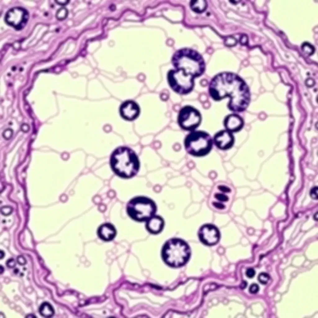
Histology
Histology is a branch of biology and medicine focused on studying tissue structure, function, and disease states at the microscopic level. It involves the examination of tissue samples from a variety of organisms, including single-celled organisms, plants, fungi, and animals, using specialized chemical stains that are tailored to highlight specific tissue features. Histopathology, a clinical application of histology, applies these techniques to analyze diseased cells and tissues, aiding in the diagnostic and prognostic assessment of various medical conditions such as cancer and multi-organ diseases. Additionally, histopathology is instrumental in identifying pathogens like bacteria, fungi, and parasites, and in detecting the presence of heavy metals and other toxins, thereby contributing to a better understanding of health conditions and their causes.
Overview
Staining in Histopathology
Numerous staining techniques were initially developed empirically for analyzing tissue sections. Classical stains like H&E and Fuchsin are suitable for the majority of diagnoses (90-95%). However, more complex diagnoses require additional differential staining methods, such as Wright-Giemsa and Schiff staining, which help reveal additional morphological criteria and functional properties. Modern techniques, including immunohistochemistry, DNA hybridization, fluorescence in situ hybridization, PCR, and flow cytometry, augment traditional histopathologic analysis.
Importance of Standardized Histological Staining
Histological and cytological diagnostics rely on the proper binding of dyes, stains, or immune probes to the key cell and tissue chemical structures that indicate the cellular architecture or pathological state. To achieve consistent and reliable results, tissue preservation and permeability are crucial. Factors like fixation type, fixation time, tissue thickness, and target accessibility influence staining effectiveness. Histopathology laboratories follow standardized fixation and staining protocols to minimize artifacts and ensure high-quality results for accurate diagnosis.