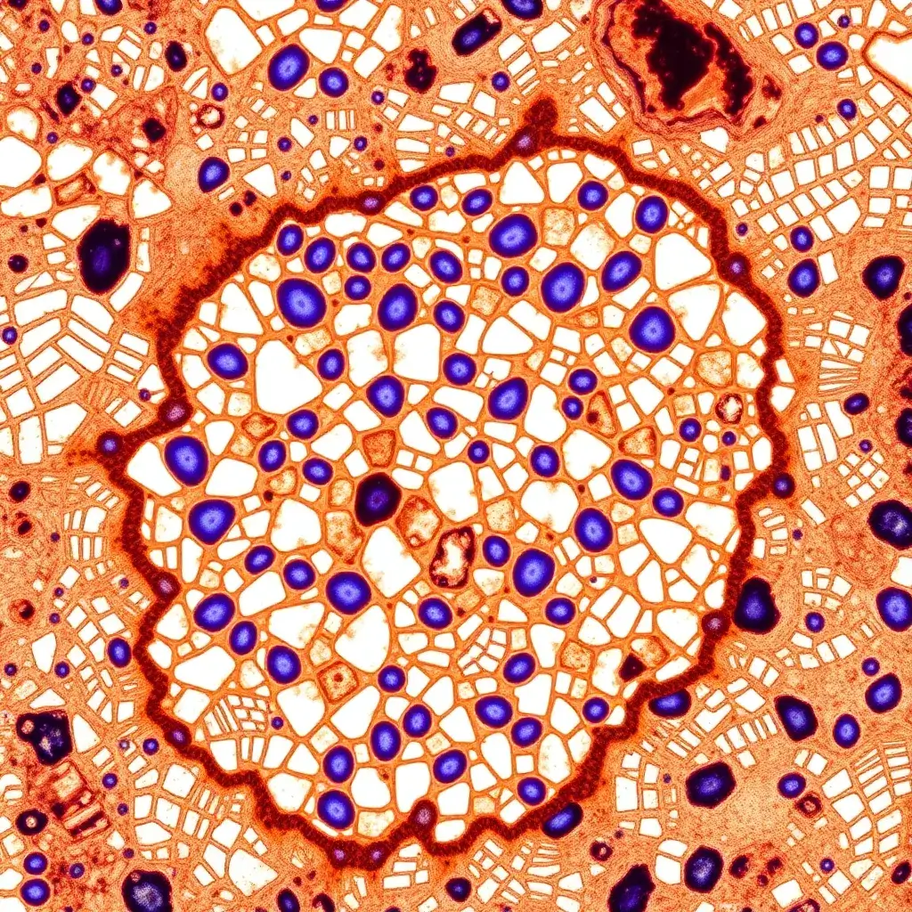
Immunohistochemistry
Diagnostic immunohistochemistry (IHC) is a group of techniques designed to detect tissue abnormalities, establish prognostic markers, and suggest potential therapeutic options. The core principle of diagnostic IHC involves an antigen-antibody binding reaction, where an antibody, conjugated with an enzyme or fluorescent dye, binds to a specific antigen within tissue samples or tissue sections, allowing the visualization of its localization and distribution. While histological staining has been a primary analytical tool for over a century, advancements in diagnostic immunohistochemistry and molecular analysis techniques have significantly transformed modern clinical pathology, enabling more precise and effective diagnoses, prognosis assessments, and the identification of targeted therapeutic strategies.
Overview
Diagnostic IHC and Clinical Pathology
Immunohistochemistry (IHC) has become a crucial analytical and diagnostic tool in general pathology laboratories, offering valuable insights into clinical diagnostics. While traditional tissue staining methods like hematoxylin and eosin (H&E) remain standard, IHC plays a significant role in detecting early cellular changes such as proliferation or apoptosis. Clinical antibodies in IHC help identify specific cell lineages by binding to marker proteins. However, IHC's sensitivity to tissue handling, preservation, and reagent quality can sometimes result in false-positive or false-negative results. To mitigate errors, clinical pathology laboratories adhere to established protocols and employ clinically validated antibodies, stains, and reagents.
Diagnostic IHC and Cancers
Cancer detection often involves a combination of histological staining and IHC to identify specific antigens in biopsy tissues. IHC provides rapid and accurate identification, particularly when traditional H&E staining proves challenging, such as in metastatic or poorly differentiated tumors. IHC is particularly useful in cases of Cancer of Unknown Primary (CUP), where the primary tumor site remains undetected despite metastatic spread. It helps clarify the diagnosis by identifying tumor origin and behavior. IHC also plays a crucial role in diagnosing cancers like adenocarcinomas (colon, breast, prostate) and skin cancers, where it complements other diagnostic methods such as microsatellite instability (MSI) testing in colon cancer diagnostics.
Diagnostic IHC and Infectious Agents
Diagnostic IHC offers rapid, morphologic differentiation of infections in tissue samples, aiding in timely clinical decisions. It is particularly effective for detecting microorganisms that are difficult to identify using routine stains, are present in low numbers, or are non-cultivable. IHC methods are used to confirm infections such as Hepatitis B, Hepatitis C, and Cytomegalovirus infections by targeting microbial DNA or RNA. It is also invaluable in detecting cutaneous infections, where traditional stains may fail. Immunofluorescence assays (IFA) have been extensively used to identify viral, bacterial, and protozoal pathogens in both human and veterinary medicine, further enhancing the capability of IHC in infectious disease diagnostics.