Flow Cytometry
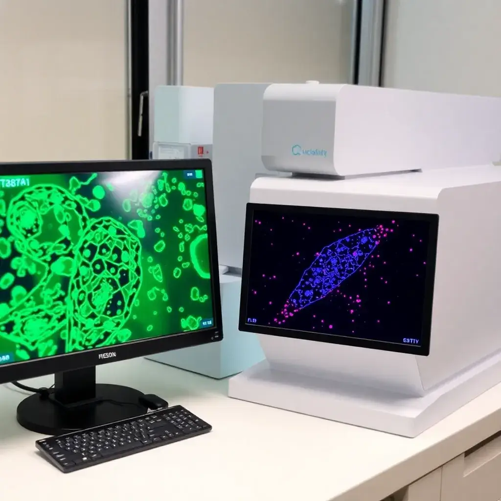
Flow cytometry is a technology that uses single or multiple lasers to provide a multi-parametric analysis of single cells. Each cell or particle is analyzed by visible light scatter or by fluorescence as cells rapidly flow past each laser. Independent of light scatter analysis, fluorescence measurements are achieved by transfection and expression of common fluorescent proteins (e.g. Green Fluorescent Protein, GFP) and staining with fluorescently conjugated antibodies or fluorescent dyes. Flow cytometry is a powerful technology that is commonly used in molecular and cellular biology research applications, including immunology, cancer biology, and disease research
Overview
Flow Cytometry Antibodies and Labels
Flow cytometry is a powerful technique used to analyze cells by using specific antibodies and labels that bind to particular cell markers, providing insights into cell types and protein expression changes. Flow cytometry antibodies are selected based on the specific cellular target, enabling researchers to analyze different aspects of cell biology. These antibodies can be labeled with a variety of different markers to facilitate detection, depending on the experimental design and the parameters under study.
Common labels include nucleic acid dyes, which bind to DNA and RNA to assist in cell cycle analysis, and cell viability dyes, which help distinguish between live and dead cells. Other types of labels include polymer dyes, quantum dots, small organic molecules, and fluorescent proteins. Antibodies are typically labeled using direct conjugation techniques, either through commercially available antibody conjugates or by using conjugation kits that allow researchers to label antibodies in the lab. In some cases, secondary antibodies that recognize the primary antibody are used for enhanced detection sensitivity.
Flow Cytometry Applications
Flow cytometry is utilized across a wide range of scientific disciplines, particularly those involving molecular biology, immunology, and cell biology. It allows for the simultaneous analysis of multiple parameters within diverse cell populations, making it a highly versatile tool. One of its most common applications is in the analysis of recombinantly expressed fluorescent proteins, which are used to investigate gene function, track cells in vivo, and study cellular behaviors. Additionally, DNA staining is frequently employed for cell cycle analysis, enabling researchers to assess the distribution of cells in different phases of the cell cycle.
Another key application of flow cytometry is the study of signal transduction pathways, where antibodies are used to detect and quantify protein expression levels, providing valuable insights into cellular responses to stimuli. Immunophenotyping is another prominent application, where fluorochrome-conjugated antibodies are used to target multiple cell surface antigens. This allows for the identification and classification of specific immune cells within heterogeneous cell populations, helping researchers to study immune responses, diseases, and therapies.
Flow Cytometry Data Analysis
Analyzing flow cytometry data typically involves identifying specific populations of cells or clusters and applying various parameters to understand cellular characteristics. Researchers often look for additional identifying features, such as the presence of specific cell markers or changes in protein expression, to gain deeper insights into the biology of the sample. Flow cytometry software plays a crucial role in this analysis, providing tools to visualize and interpret complex data sets. These programs allow for the creation of scatter plots, histograms, and other graphical representations that highlight key features of the data, enabling researchers to track changes in cellular behavior.
In addition to the general analysis, flow cytometry is often used for cell cycle analysis, where researchers can evaluate the progression of cells through different stages, such as G0/G1, S, and G2/M. Software programs can automate many of these analyses, increasing throughput and precision in research settings.
Flow Cytometry Instrumentation
Flow cytometry instrumentation is composed of several critical components, including electronics, optics, and fluidics. The fluidics system is responsible for guiding the sample through the flow cytometer, where it is exposed to the laser excitation system. The optics system consists of the laser used to excite fluorochromes and the collection optics, which include photodiodes and photomultiplier tubes (PMTs) that detect the emitted light. These detectors measure various properties of the cells, such as size, granularity, and fluorescence intensity, allowing for the characterization of individual cells within the sample.
The electronics in a flow cytometer convert the light signals from the detectors into digital data that can be analyzed and interpreted by computers. In addition to the basic flow cytometry system, more advanced technologies, such as cell sorters, can be integrated to separate specific cell populations based on their fluorescence or other markers. Imaging cytometers combine flow cytometry with fluorescence microscopy to enable more detailed imaging of cells and their environments. These advanced systems expand the capabilities of flow cytometry, making it a more powerful tool for high-throughput cell analysis and sorting.