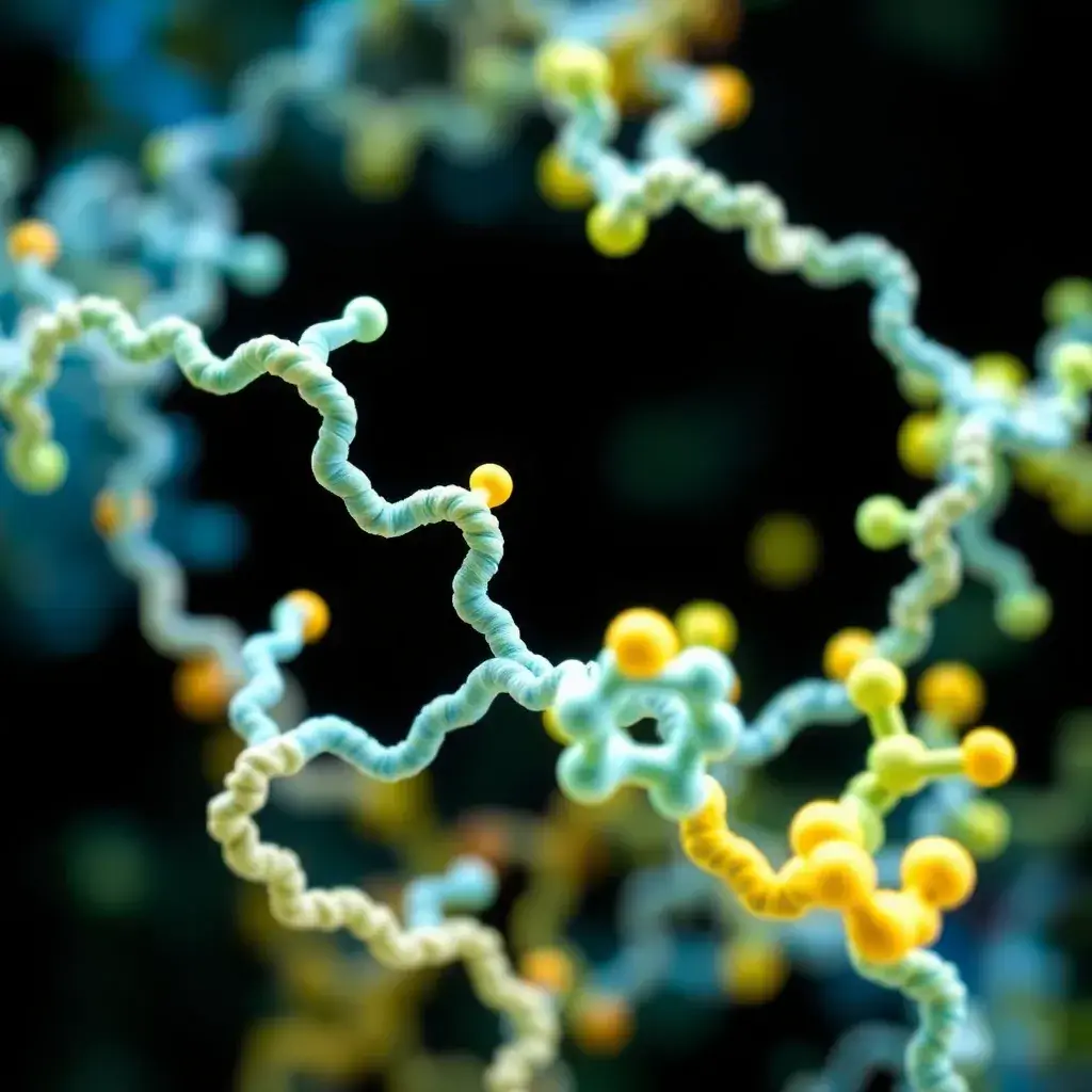
Protein Structural Analysis
The function of a protein is directly influenced by its three-dimensional structure, its interactions with other proteins, and its specific location within cells, tissues, and organs. The study of protein structure and function, known as proteomics, is conducted on a large scale and is essential for identifying protein biomarkers that are linked to specific disease states, offering potential targets for therapeutic interventions. Gaining a deeper understanding of protein structure, as well as mapping protein locations, expression levels, and their interactions, provides valuable insights that can be used to infer their biological function and role in cellular processes,
Overview
Protein Structure
Protein structure is determined by the sequence of amino acids that compose the protein and how the protein folds into more complex shapes. Protein structure can be broken down into several levels:
- Primary structure: Defined by the amino acid sequence of the protein.
- Secondary structure: Defined by local interactions of stretches of the polypeptide chain, which can form α-helices and β-sheets through hydrogen bonding interactions.
- Tertiary structure: Defines the overall three-dimensional structure of the protein.
- Quaternary structure: Defines how multiple protein subunits interact to form larger complexes.
Protein Structure Determination
The determination of three-dimensional protein structures at atomic resolution is useful in the elucidation of protein function, structure-based drug design, and molecular docking. Methods used for protein structure determination include:
- NMR: Nuclear magnetic resonance (NMR) spectroscopy is used to obtain information about the structure and dynamics of proteins. In NMR, the spatial location of atoms is determined by their chemical shifts. For protein NMR, proteins are typically labeled with stable isotopes (15N, 13C, 2H) to enhance sensitivity and facilitate structural deconvolution. Isotopic labels are typically introduced by supplying isotopically labeled nutrients in the growth medium during protein expression.
- X-ray Crystallography: Protein X-ray crystallography can be used to obtain the three-dimensional structure of proteins through X-ray diffraction of crystallized proteins. Crystals are grown by seeding highly concentrated protein in solutions that promote precipitation, with ordered protein crystals forming under suitable conditions. X-rays are aimed at the protein crystal, which scatters the X-rays onto an electronic detector or film. The crystals are rotated to capture diffraction in three dimensions, enabling calculation of the position of each atom in the crystallized molecule by Fourier Transform.
Protein Mapping
Mapping of the location and expression level of proteins in specific cells, tissues, and organs aids in the functional study of the proteome. Spatial distribution of proteins is key to protein function, with improper localization or expression triggering various disease states. Key aspects of protein mapping include:
- Human Protein Atlas: A resource providing comprehensive data on protein localization and expression levels, supporting biomarker discovery and understanding disease pathology.
- Interactome Mapping: Mapping of the interactome helps define the molecular interactions that occur on a cellular level, assisting in the understanding of protein function and providing valuable potential drug targets for disease.