S5000
Johannes Lieder Animal Cell Set
Manufacturer: Johannes Lieder
Select a Size
| Pack Size | SKU | Availability | Price |
|---|---|---|---|
| Each of 1 | S5000-Each-of-1 | In Stock | ₹ 26,700.00 |
S5000 - Each of 1
In Stock
Quantity
1
Base Price: ₹ 26,700.00
GST (18%): ₹ 4,806.00
Total Price: ₹ 31,506.00
Type
The Animal Cell
Quantity
12
Related Products


Description
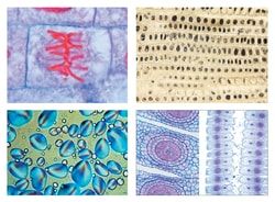
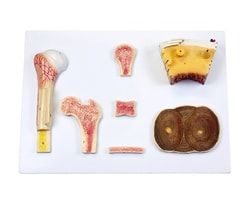
- Set contains 12 slides of animal cytology with depictured accompanying brochure
- Epithelium, muscle, bone, nerve, internal organs, sex organs and chromosomes are all represented in this slide set
- It shows a fine cross section of tissue types useful for classroom microscope study
- Includes: Squamous epithelium, isolated cells from human mouth
- Nuclei and cytoplasm are shown Striated muscle l.s
- showing nuclei, striations, myofibrils Compact bone and hyaline cartilage t.s., two sections on one slide for comparison Nerve fibres isolated, fixed and stained by osmic acid to show myeline sheaths and Ranvier’s nodes Liver of Salamandra t.s., showing simple animal cells with cellular membranes, nuclei, and cytoplasm Kidney of mouse, t.s
- vital stained with trypanblue to demonstrate the storage of epithelial cells Ovary of cat, t.s
- showing primary, secondary, and Graafian follicles Testis of frog, t.s
- showing spermatogenesis
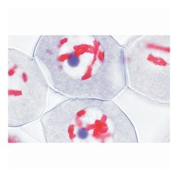
- Spermatogonia, spermatocytes, spermatids, and mature spermatozoa Salamandra larva, t.s
- of skin and other organs selected to show cell division (mitosis) in various stages Uteri of Ascaris megalocephala, t.s
- iron hematoxyline stained to show details of meiosis with chromosomes and nuclear spindles Salivary gland of Chironomus larva
- Giant chromosomes showing large chromomeres
- Stained for DNA after Feulgen Ova from Psammechinus (sea urchin)
- Unfertilized ova, fertilized ova, early cleavage stages