BDB550803
BD BrdU In-Situ Detection Kit
Manufacturer: BD
Select a Size
| Pack Size | SKU | Availability | Price |
|---|---|---|---|
| Each of 1 | BDB550803-Each-of-1 | In Stock | ₹ 40,584.00 |
BDB550803 - Each of 1
In Stock
Quantity
1
Base Price: ₹ 40,584.00
GST (18%): ₹ 7,305.12
Total Price: ₹ 47,889.12
Quantity
1 Kit
Related Products

Description
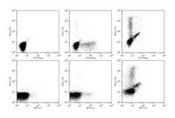
- Cellular incorporation of BrdU during S phase can be readily detected by anti-BrdU-specific antibodies following membrane permeabilization
- Subsequent quantitation of cells positive for BrdU is possible using routine methods for cell analysis Measures labeling indices of different cell populations Improved specific staining with minimal background Designed antigen retrieval solution unmask antigenic sites and preserve tissue morphology Enables study of proliferation state of phenotypically defined cells within the micro-environment of tissues Reaction: 50 slides Flow Cytometry, Immunohistochemistry