50-112-8705
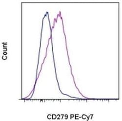
CD279 (PD-1) Monoclonal Antibody (J43), PE-Cyanine7, eBioscience™, Invitrogen™
Manufacturer: Fischer Scientific
Select a Size
| Pack Size | SKU | Availability | Price |
|---|---|---|---|
| Each of 1 | 50-112-8705-Each-of-1 | In Stock | ₹ 33,553.00 |
50-112-8705 - Each of 1
In Stock
Quantity
1
Base Price: ₹ 33,553.00
GST (18%): ₹ 6,039.54
Total Price: ₹ 39,592.54
Antigen
CD279 (PD-1)
Classification
Monoclonal
Concentration
0.2 mg/mL
Formulation
PBS with 0.09% sodium azide; pH 7.2
Gene Accession No.
Q02242
Gene Symbols
Pdcd1
Purification Method
Affinity chromatography
Regulatory Status
RUO
Gene ID (Entrez)
18566
Content And Storage
4° C, store in dark, DO NOT FREEZE!
Form
Liquid
Applications
Flow Cytometry
Clone
J43
Conjugate
PE-Cyanine7
Gene
Pdcd1
Gene Alias
CD279; EGK_05005; hPD1; hPD-1; hPD-l; hSLE1; Ly101; mPD-1; PD1; PD-1; Pdc1; Pdcd1; programmed cell death 1; programmed cell death 1 protein; programmed cell death protein 1; programmed cell death protein 1-like; programmed death 1; Protein PD1; protein PD-1; sCD279; SLEB2; soluble CD279; systemic lupus erythematosus susceptibility 2
Host Species
Armenian Hamster
Quantity
100 μg
Primary or Secondary
Primary
Target Species
Mouse
Product Type
Antibody
Isotype
IgG
Description
- Description: The J43 monoclonal antibody reacts with mouse PD-1 (programmed death-1), a 55 kDa member of the Ig superfamily
- PD-1 contains the immunoreceptor tyrosine-based inhibitory motif (ITIM) and plays a key role in peripheral tolerance and autoimmune disease in mice
- PD-1 is expressed mainly on activated T and B lymphocytes
- Two novel B7 Family members have been identified as PD-1 ligands, PD-L1 (B7-H1) and PD-L2 (B7-DC)
- Evidence reported to date suggests overlapping functions for these ligands and their constitutive expression on some normal tissues and upregulation on activated antigen-presenting cells
- It is reported that J43 inhibits the binding of mouse PD-L1-Ig and mouse PD-L2-Ig to PD-1/BHK transfected cells
- When administrated in vivo, both intact and Fab of J43 are reported to enhance contact hypersensitivity and exacerbate acute GVHD similar to transfer of PD-1-deficient cells
- Injection of J43 also exacerbates EAE and NOD diabetes as do specific antibodies to mouse PD-L1 and PD-L2
- Applications Reported: This J43 antibody has been reported for use in flow cytometric analysis
- Applications Tested: This J43 antibody has been tested by flow cytometric analysis of stimulated mouse splenocytes
- This can be used at less than or equal to 1 μg per test
- A test is defined as the amount (μg) of antibody that will stain a cell sample in a final volume of 100 μL
- Cell-mediated immune responses are initiated by T lymphocytes that are themselves stimulated by cognate peptides bound to MHC molecules on antig en-presenting cells (APC)
- T-cell activation is generally self-limited as activated T cells express receptors such as PD-1 (also known as PDCD-1) that mediate inhibitory signals from the APC
- PD-1 can bind two different but related ligands, PDL-1 and PDL-2
- Upon binding to either of these ligands, signals generated by PD-1 inhibit the activation of the immune response in the absence of "edanger signals"e such as LPS or other molecules associated with bacteria or other pathogens
- Evidence for this is seen in PD1-null mice who exhibit hyperactivated immune systems and autoimmune diseases
- Despite its predicted molecular weight, PD-1 often migrates at higher molecular weight in SDS-PAGE.
Compare Similar Items
Show Difference
Antigen: CD279 (PD-1)
Classification: Monoclonal
Concentration: 0.2 mg/mL
Formulation: PBS with 0.09% sodium azide; pH 7.2
Gene Accession No.: Q02242
Gene Symbols: Pdcd1
Purification Method: Affinity chromatography
Regulatory Status: RUO
Gene ID (Entrez): 18566
Content And Storage: 4° C, store in dark, DO NOT FREEZE!
Form: Liquid
Applications: Flow Cytometry
Clone: J43
Conjugate: PE-Cyanine7
Gene: Pdcd1
Gene Alias: CD279; EGK_05005; hPD1; hPD-1; hPD-l; hSLE1; Ly101; mPD-1; PD1; PD-1; Pdc1; Pdcd1; programmed cell death 1; programmed cell death 1 protein; programmed cell death protein 1; programmed cell death protein 1-like; programmed death 1; Protein PD1; protein PD-1; sCD279; SLEB2; soluble CD279; systemic lupus erythematosus susceptibility 2
Host Species: Armenian Hamster
Quantity: 100 μg
Primary or Secondary: Primary
Target Species: Mouse
Product Type: Antibody
Isotype: IgG
Antigen:
CD279 (PD-1)
Classification:
Monoclonal
Concentration:
0.2 mg/mL
Formulation:
PBS with 0.09% sodium azide; pH 7.2
Gene Accession No.:
Q02242
Gene Symbols:
Pdcd1
Purification Method:
Affinity chromatography
Regulatory Status:
RUO
Gene ID (Entrez):
18566
Content And Storage:
4° C, store in dark, DO NOT FREEZE!
Form:
Liquid
Applications:
Flow Cytometry
Clone:
J43
Conjugate:
PE-Cyanine7
Gene:
Pdcd1
Gene Alias:
CD279; EGK_05005; hPD1; hPD-1; hPD-l; hSLE1; Ly101; mPD-1; PD1; PD-1; Pdc1; Pdcd1; programmed cell death 1; programmed cell death 1 protein; programmed cell death protein 1; programmed cell death protein 1-like; programmed death 1; Protein PD1; protein PD-1; sCD279; SLEB2; soluble CD279; systemic lupus erythematosus susceptibility 2
Host Species:
Armenian Hamster
Quantity:
100 μg
Primary or Secondary:
Primary
Target Species:
Mouse
Product Type:
Antibody
Isotype:
IgG
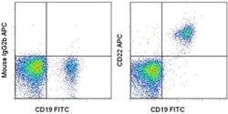
Antigen: CD22
Classification: Monoclonal
Concentration: 5 μL/Test
Formulation: PBS with 0.2% BSA and 0.09% sodium azide; pH 7.2
Gene Accession No.: P20273
Gene Symbols: CD22
Purification Method: Affinity chromatography
Regulatory Status: RUO
Gene ID (Entrez): 933
Content And Storage: 4° C, store in dark, DO NOT FREEZE!
Form: Liquid
Applications: Flow Cytometry
Clone: S-HCL-1
Conjugate: APC
Gene: CD22
Gene Alias: A530093D23; B-cell receptor CD22; BL-CAM; B-lymphocyte cell adhesion molecule; B-lymphocyte cell adhesion molecule (BL-CAM); Cd22; CD22 antigen; CD22 molecule; FLJ22814; Lectin 2; Leu-14; Lyb8; Lyb-8; MGC130020; sialic acid binding Ig-like lectin 2; sialic acid-binding Ig-like lectin 2; Sialic acid-binding Ig-like lectin 2 (Siglec-2); Siglec2; Siglec-2; T-cell surface antigen Leu-14
Host Species: Mouse
Quantity: 100 Tests
Primary or Secondary: Primary
Target Species: Human
Product Type: Antibody
Isotype: IgG2b κ
Antigen:
CD22
Classification:
Monoclonal
Concentration:
5 μL/Test
Formulation:
PBS with 0.2% BSA and 0.09% sodium azide; pH 7.2
Gene Accession No.:
P20273
Gene Symbols:
CD22
Purification Method:
Affinity chromatography
Regulatory Status:
RUO
Gene ID (Entrez):
933
Content And Storage:
4° C, store in dark, DO NOT FREEZE!
Form:
Liquid
Applications:
Flow Cytometry
Clone:
S-HCL-1
Conjugate:
APC
Gene:
CD22
Gene Alias:
A530093D23; B-cell receptor CD22; BL-CAM; B-lymphocyte cell adhesion molecule; B-lymphocyte cell adhesion molecule (BL-CAM); Cd22; CD22 antigen; CD22 molecule; FLJ22814; Lectin 2; Leu-14; Lyb8; Lyb-8; MGC130020; sialic acid binding Ig-like lectin 2; sialic acid-binding Ig-like lectin 2; Sialic acid-binding Ig-like lectin 2 (Siglec-2); Siglec2; Siglec-2; T-cell surface antigen Leu-14
Host Species:
Mouse
Quantity:
100 Tests
Primary or Secondary:
Primary
Target Species:
Human
Product Type:
Antibody
Isotype:
IgG2b κ
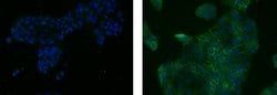
Antigen: CD324 (E-Cadherin)
Classification: Monoclonal
Concentration: 0.5 mg/mL
Formulation: PBS with 0.09% sodium azide; pH 7.2
Gene Accession No.: F1PAA9, P09803, P12830
Gene Symbols: CDH1
Purification Method: Affinity chromatography
Regulatory Status: RUO
Gene ID (Entrez): 12550, 442858, 999
Content And Storage: 4° C, store in dark, DO NOT FREEZE!
Form: Liquid
Applications: Flow Cytometry, Immunocytochemistry, Immunohistochemistry (Frozen)
Clone: DECMA-1
Conjugate: Alexa Fluor 488
Gene: CDH1
Gene Alias: AA960649; ARC-1; cadherin 1; cadherin 1, E-cadherin (epithelial); cadherin 1, epithelial; cadherin 1, type 1; cadherin 1, type 1, E-cadherin (epithelial); cadherin e; cadherin-1; cadherin-E; calcium-dependent adhesion protein, epithelial; CAM 120/80; CD324; CDH1; CDHE; cell adhesion molecule; cell-CAM 120/80; ECAD; E-cad; E-Cad/CTF1; E-Cad/CTF2; E-Cad/CTF3; Ecadherin; E-cadherin; E-cadherin 1; E-cadherin precursor; Epithelial cadherin; hab; half baked; LCAM; L-CAM; Um; UVO; Uvomorulin
Host Species: Rat
Quantity: 100 μg
Primary or Secondary: Primary
Target Species: Canine, Human, Mouse
Product Type: Antibody
Isotype: IgG1 κ
Antigen:
CD324 (E-Cadherin)
Classification:
Monoclonal
Concentration:
0.5 mg/mL
Formulation:
PBS with 0.09% sodium azide; pH 7.2
Gene Accession No.:
F1PAA9, P09803, P12830
Gene Symbols:
CDH1
Purification Method:
Affinity chromatography
Regulatory Status:
RUO
Gene ID (Entrez):
12550, 442858, 999
Content And Storage:
4° C, store in dark, DO NOT FREEZE!
Form:
Liquid
Applications:
Flow Cytometry, Immunocytochemistry, Immunohistochemistry (Frozen)
Clone:
DECMA-1
Conjugate:
Alexa Fluor 488
Gene:
CDH1
Gene Alias:
AA960649; ARC-1; cadherin 1; cadherin 1, E-cadherin (epithelial); cadherin 1, epithelial; cadherin 1, type 1; cadherin 1, type 1, E-cadherin (epithelial); cadherin e; cadherin-1; cadherin-E; calcium-dependent adhesion protein, epithelial; CAM 120/80; CD324; CDH1; CDHE; cell adhesion molecule; cell-CAM 120/80; ECAD; E-cad; E-Cad/CTF1; E-Cad/CTF2; E-Cad/CTF3; Ecadherin; E-cadherin; E-cadherin 1; E-cadherin precursor; Epithelial cadherin; hab; half baked; LCAM; L-CAM; Um; UVO; Uvomorulin
Host Species:
Rat
Quantity:
100 μg
Primary or Secondary:
Primary
Target Species:
Canine, Human, Mouse
Product Type:
Antibody
Isotype:
IgG1 κ

Antigen: CD56 (NCAM)
Classification: Monoclonal
Concentration: 5 μL/Test
Formulation: PBS with 0.2% BSA and 0.09% sodium azide; pH 7.2
Gene Accession No.: P13591
Gene Symbols: Ncam1
Purification Method: Affinity chromatography
Regulatory Status: RUO
Gene ID (Entrez): 4684
Content And Storage: 4° C, store in dark, DO NOT FREEZE!
Form: Liquid
Applications: Flow Cytometry
Clone: CMSSB
Conjugate: APC
Gene: Ncam1
Gene Alias: adhesion molecule; antigen recognized by monoclonal antibody 5.1H11; Cd56; CD-56; CD56 120 kDa GPI-linked isoform; CD56 140 kDa isoform; CD56 140 kDa VASE isoform; E NCAM; E-NCAM; MSK39; N CAM1; NCAM; N-CAM; Ncam1; N-CAM-1; NCAM-1; NCAMC; NCAM-C; neural cell adhesion molecule; Neural cell adhesion molecule 1; neural cell adhesion molecule, NCAM; sCD56; sNCAM; soluble CD56; soluble NCAM
Host Species: Mouse
Quantity: 100 Tests
Primary or Secondary: Primary
Target Species: Human
Product Type: Antibody
Isotype: IgG1 κ
Antigen:
CD56 (NCAM)
Classification:
Monoclonal
Concentration:
5 μL/Test
Formulation:
PBS with 0.2% BSA and 0.09% sodium azide; pH 7.2
Gene Accession No.:
P13591
Gene Symbols:
Ncam1
Purification Method:
Affinity chromatography
Regulatory Status:
RUO
Gene ID (Entrez):
4684
Content And Storage:
4° C, store in dark, DO NOT FREEZE!
Form:
Liquid
Applications:
Flow Cytometry
Clone:
CMSSB
Conjugate:
APC
Gene:
Ncam1
Gene Alias:
adhesion molecule; antigen recognized by monoclonal antibody 5.1H11; Cd56; CD-56; CD56 120 kDa GPI-linked isoform; CD56 140 kDa isoform; CD56 140 kDa VASE isoform; E NCAM; E-NCAM; MSK39; N CAM1; NCAM; N-CAM; Ncam1; N-CAM-1; NCAM-1; NCAMC; NCAM-C; neural cell adhesion molecule; Neural cell adhesion molecule 1; neural cell adhesion molecule, NCAM; sCD56; sNCAM; soluble CD56; soluble NCAM
Host Species:
Mouse
Quantity:
100 Tests
Primary or Secondary:
Primary
Target Species:
Human
Product Type:
Antibody
Isotype:
IgG1 κ