Ki-67 Monoclonal Antibody (20Raj1), FITC, eBioscience™, Invitrogen™
Manufacturer: Fischer Scientific
Select a Size
| Pack Size | SKU | Availability | Price |
|---|---|---|---|
| Each of 1 | 50-112-8738-Each-of-1 | In Stock | ₹ 32,841.00 |
50-112-8738 - Each of 1
In Stock
Quantity
1
Base Price: ₹ 32,841.00
GST (18%): ₹ 5,911.38
Total Price: ₹ 38,752.38
Antigen
Ki-67
Classification
Monoclonal
Concentration
5 μL/Test
Formulation
PBS with 0.2% BSA and 0.09% sodium azide; pH 7.2
Gene Accession No.
P46013
Gene Symbols
Mki67
Purification Method
Affinity chromatography
Regulatory Status
RUO
Gene ID (Entrez)
100686578, 4288
Content And Storage
4° C, store in dark, DO NOT FREEZE!
Form
Liquid
Applications
Flow Cytometry, Immunocytochemistry, Immunohistochemistry
Clone
20Raj1
Conjugate
FITC
Gene
Mki67
Gene Alias
antigen identified by monoclonal antibody Ki 67; antigen identified by monoclonal antibody Ki-67; Antigen identified by monoclonal antibody Ki-67 homolog; Antigen KI-67; Antigen KI-67 homolog; antigen KI-67; proliferation marker protein Ki-67; antigen KI-67-like; cb31; D630048A14Rik; I79_022666; Ki67; Ki-67; KIA; LOW QUALITY PROTEIN: proliferation marker protein Ki-67; marker of proliferation Ki-67; MIB-; MIB-1; Mki67; PPP1R105; Proliferation marker protein Ki-67; proliferation-related Ki-67 antigen; protein phosphatase 1, regulatory subunit 105; RP11-380J17.2; sb:cb31; si:ch211-250b22.7; unnamed protein product; wu:fa11g09; wu:fb57a07; wu:fi14e05
Host Species
Mouse
Quantity
100 Tests
Primary or Secondary
Primary
Target Species
Canine, Human
Product Type
Antibody
Isotype
IgG1 κ
Description
- Description: The monoclonal antibody 20Raj1 recognizes the human Ki-67 protein
- Two isoforms of Ki-67 exist, a 345 and 395 kDa form that are expressed in dividing cells
- Ki-67 is expressed in all cell types and is detectable during active phases of the cell cycle (G1, S, G2, and mitosis) but is absent from resting cells (G0)
- During interphase, Ki-67 expression is localized to the nucleus but redistributes to the chromosomes during mitosis and has specifically been found to associate with heterochromatin-bound proteins such as chromobox protein homolog 3 (CBX3)
- In studies of tumor cells, Ki-67 expression has been used as a marker for determining the fraction of proliferating cells within a given population of tumor cells
- This monoclonal antibody 20Raj1 recognizes canine Ki-67
- Applications Reported: This 20Raj1 antibody has been reported for use in intracellular staining followed by flow cytometric analysis, immunohistochemical staining, and immunocytochemistry
- Applications Tested: This 20Raj1 antibody has been pre-titrated and tested by intracellular staining and flow cytometric analysis of stimulated normal human peripheral blood cells using the Foxp3/Transcription Factor Staining Buffer Set (cat
- 00-5523) and protocol
- Please see Best Protocols Section (Staining intracellular Antigens for Flow Cytometry) for staining protocol (refer to Protocol B: One-step protocol for intracellular (nuclear) proteins)
- This can be used at 5 μL (0.06 μg) per test
- Ki-67 is a nuclear protein that is expressed during various stages in the cell cycle, particularly during late G1, S, G2, and M phases
- The protein has a forkhead associated domain (FHA) through which it associates with euchromatin at the perichromosomal layer, the centromeric heterochromatin, and the nucleolus
- Ki-67 is shown to have a cell cycle dependent topographical distribution with perinucleolar expression at G1, expression in the nuclear matrix at G2, and expression on the chromosomes during M phase
- Ki-67 is commonly used as a proliferation marker because it is not detected in G0 cells, but increases steadily from G1 through mitosis
- Ki-67 antibodies are useful in establishing the cell growing fraction in neoplasms
- In neoplastic tissues, the prognostic value is comparable to the tritiated thymidine-labelling index
- The correlation between low Ki-67 index and histologically low-grade tumors is strong
- Ki-67 is routinely used as a neuronal marker of cell cycling and proliferation.
Compare Similar Items
Show Difference

Antigen: Ki-67
Classification: Monoclonal
Concentration: 5 μL/Test
Formulation: PBS with 0.2% BSA and 0.09% sodium azide; pH 7.2
Gene Accession No.: P46013
Gene Symbols: Mki67
Purification Method: Affinity chromatography
Regulatory Status: RUO
Gene ID (Entrez): 100686578, 4288
Content And Storage: 4° C, store in dark, DO NOT FREEZE!
Form: Liquid
Applications: Flow Cytometry, Immunocytochemistry, Immunohistochemistry
Clone: 20Raj1
Conjugate: FITC
Gene: Mki67
Gene Alias: antigen identified by monoclonal antibody Ki 67; antigen identified by monoclonal antibody Ki-67; Antigen identified by monoclonal antibody Ki-67 homolog; Antigen KI-67; Antigen KI-67 homolog; antigen KI-67; proliferation marker protein Ki-67; antigen KI-67-like; cb31; D630048A14Rik; I79_022666; Ki67; Ki-67; KIA; LOW QUALITY PROTEIN: proliferation marker protein Ki-67; marker of proliferation Ki-67; MIB-; MIB-1; Mki67; PPP1R105; Proliferation marker protein Ki-67; proliferation-related Ki-67 antigen; protein phosphatase 1, regulatory subunit 105; RP11-380J17.2; sb:cb31; si:ch211-250b22.7; unnamed protein product; wu:fa11g09; wu:fb57a07; wu:fi14e05
Host Species: Mouse
Quantity: 100 Tests
Primary or Secondary: Primary
Target Species: Canine, Human
Product Type: Antibody
Isotype: IgG1 κ
Antigen: CD279 (PD-1)
Classification: Monoclonal
Concentration: 0.5 mg/mL
Formulation: PBS with 0.09% sodium azide; pH 7.2
Gene Accession No.: Q02242
Gene Symbols: Pdcd1
Purification Method: Affinity chromatography
Regulatory Status: RUO
Gene ID (Entrez): 18566
Content And Storage: 4° C, store in dark, DO NOT FREEZE!
Form: Liquid
Applications: Flow Cytometry
Clone: J43
Conjugate: Biotin
Gene: Pdcd1
Gene Alias: CD279; EGK_05005; hPD1; hPD-1; hPD-l; hSLE1; Ly101; mPD-1; PD1; PD-1; Pdc1; Pdcd1; programmed cell death 1; programmed cell death 1 protein; programmed cell death protein 1; programmed cell death protein 1-like; programmed death 1; Protein PD1; protein PD-1; sCD279; SLEB2; soluble CD279; systemic lupus erythematosus susceptibility 2
Host Species: Armenian Hamster
Quantity: 100 μg
Primary or Secondary: Primary
Target Species: Mouse
Product Type: Antibody
Isotype: IgG
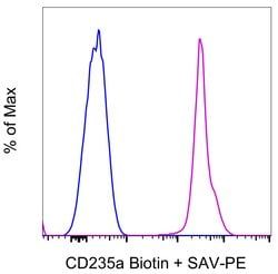
Antigen: CD235a (Glycophorin A)
Classification: Monoclonal
Concentration: 0.5 mg/mL
Formulation: PBS with 0.09% sodium azide; pH 7.2
Gene Accession No.: P02724
Gene Symbols: GYPA
Purification Method: Affinity chromatography
Regulatory Status: RUO
Gene ID (Entrez): 2993
Content And Storage: 4° C, store in dark, DO NOT FREEZE!
Form: Liquid
Applications: Flow Cytometry
Clone: HIR2 (GA-R2)
Conjugate: Biotin
Gene: GYPA
Gene Alias: AI853584; CD235; CD235a; erythroid-lineage-specific membrane sialoglycoprotein; glycophorin A; glycophorin A (MN blood group); glycophorin A (MNS blood group); glycophorin A, GPA; glycophorin Erik; glycophorin MiI; glycophorin MiV; glycophorin SAT; glycophorin Sta type C; glycophorin-A; GPA; GPErik; GPSAT; Gypa; HGNC:4702; HGpMiV; HGpMiX; HGpMiXI; HGpSta(C); HGpStaC; Mi.V glycoprotein; Mi.V glycoprotein (24 AA); MN; MN sialoglycoprotein; MNS; PAS-2; recombinant glycophorin A-B Miltenberger-DR; sialoglycoprotein alpha
Host Species: Mouse
Quantity: 100 μg
Primary or Secondary: Primary
Target Species: Human
Product Type: Antibody
Isotype: IgG2b κ
