50-112-9000
CD279 (PD-1) Monoclonal Antibody (J43), APC-eFluor™ 780, eBioscience™, Invitrogen™
Manufacturer: Fischer Scientific
Select a Size
| Pack Size | SKU | Availability | Price |
|---|---|---|---|
| Each of 1 | 50-112-9000-Each-of-1 | In Stock | ₹ 35,422.00 |
50-112-9000 - Each of 1
In Stock
Quantity
1
Base Price: ₹ 35,422.00
GST (18%): ₹ 6,375.96
Total Price: ₹ 41,797.96
Antigen
CD279 (PD-1)
Classification
Monoclonal
Concentration
0.2 mg/mL
Formulation
PBS with 0.09% sodium azide; pH 7.2
Gene Accession No.
Q02242
Gene Symbols
Pdcd1
Purification Method
Affinity chromatography
Regulatory Status
RUO
Gene ID (Entrez)
18566
Content And Storage
4° C, store in dark, DO NOT FREEZE!
Form
Liquid
Applications
Flow Cytometry
Clone
J43
Conjugate
APC-eFluor 780
Gene
Pdcd1
Gene Alias
CD279; EGK_05005; hPD1; hPD-1; hPD-l; hSLE1; Ly101; mPD-1; PD1; PD-1; Pdc1; Pdcd1; programmed cell death 1; programmed cell death 1 protein; programmed cell death protein 1; programmed cell death protein 1-like; programmed death 1; Protein PD1; protein PD-1; sCD279; SLEB2; soluble CD279; systemic lupus erythematosus susceptibility 2
Host Species
Armenian Hamster
Quantity
100 μg
Primary or Secondary
Primary
Target Species
Mouse
Product Type
Antibody
Isotype
IgG
Description
- Description: The J43 monoclonal antibody reacts with mouse PD-1 (programmed death-1), a 55 kDa member of the Ig superfamily
- PD-1 contains the immunoreceptor tyrosine-based inhibitory motif (ITIM) and plays a key role in peripheral tolerance and autoimmune disease in mice
- PD-1 is expressed mainly on activated T and B lymphocytes
- Two novel B7 Family members have been identified as PD-1 ligands, PD-L1 (B7-H1) and PD-L2 (B7-DC)
- Evidence reported to date suggests overlapping functions for these ligands and their constitutive expression on some normal tissues and upregulation on activated antigen-presenting cells
- It is reported that J43 inhibits the binding of mouse PD-L1-Ig and mouse PD-L2-Ig to PD-1/BHK transfected cells
- When administrated in vivo, both intact and Fab of J43 are reported to enhance contact hypersensitivity and exacerbate acute GVHD similar to transfer of PD-1-deficient cells
- Injection of J43 also exacerbates EAE and NOD diabetes as do specific antibodies to mouse PD-L1 and PD-L2
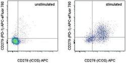
- Applications Reported: This J43 antibody has been reported for use in flow cytometric analysis
- Applications Tested: This J43 antibody has been tested by flow cytometric analysis of stimulated mouse splenocytes
- This can be used at less than or equal to 0.25 μg per test
- A test is defined as the amount (μg) of antibody that will stain a cell sample in a final volume of 100 μL
- Cell-mediated immune responses are initiated by T lymphocytes that are themselves stimulated by cognate peptides bound to MHC molecules on antig en-presenting cells (APC)
- T-cell activation is generally self-limited as activated T cells express receptors such as PD-1 (also known as PDCD-1) that mediate inhibitory signals from the APC
- PD-1 can bind two different but related ligands, PDL-1 and PDL-2
- Upon binding to either of these ligands, signals generated by PD-1 inhibit the activation of the immune response in the absence of "edanger signals"e such as LPS or other molecules associated with bacteria or other pathogens
- Evidence for this is seen in PD1-null mice who exhibit hyperactivated immune systems and autoimmune diseases
- Despite its predicted molecular weight, PD-1 often migrates at higher molecular weight in SDS-PAGE.
Compare Similar Items
Show Difference
Antigen: CD279 (PD-1)
Classification: Monoclonal
Concentration: 0.2 mg/mL
Formulation: PBS with 0.09% sodium azide; pH 7.2
Gene Accession No.: Q02242
Gene Symbols: Pdcd1
Purification Method: Affinity chromatography
Regulatory Status: RUO
Gene ID (Entrez): 18566
Content And Storage: 4° C, store in dark, DO NOT FREEZE!
Form: Liquid
Applications: Flow Cytometry
Clone: J43
Conjugate: APC-eFluor 780
Gene: Pdcd1
Gene Alias: CD279; EGK_05005; hPD1; hPD-1; hPD-l; hSLE1; Ly101; mPD-1; PD1; PD-1; Pdc1; Pdcd1; programmed cell death 1; programmed cell death 1 protein; programmed cell death protein 1; programmed cell death protein 1-like; programmed death 1; Protein PD1; protein PD-1; sCD279; SLEB2; soluble CD279; systemic lupus erythematosus susceptibility 2
Host Species: Armenian Hamster
Quantity: 100 μg
Primary or Secondary: Primary
Target Species: Mouse
Product Type: Antibody
Isotype: IgG
Antigen:
CD279 (PD-1)
Classification:
Monoclonal
Concentration:
0.2 mg/mL
Formulation:
PBS with 0.09% sodium azide; pH 7.2
Gene Accession No.:
Q02242
Gene Symbols:
Pdcd1
Purification Method:
Affinity chromatography
Regulatory Status:
RUO
Gene ID (Entrez):
18566
Content And Storage:
4° C, store in dark, DO NOT FREEZE!
Form:
Liquid
Applications:
Flow Cytometry
Clone:
J43
Conjugate:
APC-eFluor 780
Gene:
Pdcd1
Gene Alias:
CD279; EGK_05005; hPD1; hPD-1; hPD-l; hSLE1; Ly101; mPD-1; PD1; PD-1; Pdc1; Pdcd1; programmed cell death 1; programmed cell death 1 protein; programmed cell death protein 1; programmed cell death protein 1-like; programmed death 1; Protein PD1; protein PD-1; sCD279; SLEB2; soluble CD279; systemic lupus erythematosus susceptibility 2
Host Species:
Armenian Hamster
Quantity:
100 μg
Primary or Secondary:
Primary
Target Species:
Mouse
Product Type:
Antibody
Isotype:
IgG
Antigen: HLA-E
Classification: Monoclonal
Concentration: 1 mg/mL
Formulation: PBS with no preservative; pH 7.2
Gene Accession No.: P13747
Gene Symbols: HLA-E
Purification Method: Affinity chromatography
Regulatory Status: RUO
Gene ID (Entrez): 3133
Content And Storage: 4° C
Form: Liquid
Applications: Flow Cytometry, Functional Assay
Clone: 3D12HLA-E
Conjugate: Functional Grade
Gene: HLA-E
Gene Alias: A29 protein; antigen presenting molecule; Aw-33; Aw-74; DAMA-277I14.1; DAQB-90C11.16; DKFZp686P19218; EA1.2; EA2.1; FLJ26655; HLA A; HLA class I antigen; HLA class I histocompatibility antigen, A alpha chain; HLA class I histocompatibility antigen, A-1 alpha chain; HLA class I histocompatibility antigen, A-30 alpha chain; HLA class I histocompatibility antigen, A-33 alpha chain; HLA class I histocompatibility antigen, A-74 alpha chain; HLA class I histocompatibility antigen, alpha chain E; HLA class I histocompatibility antigen, E alpha chain; HLA locus; HLA-6.2; HLAA; HLA-A; HLA-A locus alpha 2 domain; HLA-A null; HLA-A11; HLA-A33; HLA-Aw33.1; HLA-DQB1; HLA-DRB1; HLAE; HLA-E; Human leukocyte antigen A; leukocyte antigen class I-A; lymphocyte antigen; major histocompatibility complex, class I, A; major histocompatibility complex, class I, E; MCH class I antigen; MHC; MHC 1; MHC class 1 antigen; MHC class I anti; MHC Class I antigen; MHC class I antigen A*30; MHC class I antigen A*33; MHC class I antigen A*74; MHC class I antigen E; MHC class I antigen heavy chain; MHC class I antigen HLA-A heavy chain; MHC class I antigen HLA-A33; MHC class I antigen null protein; MHC class I antigene; MHC class II antigen; MHC classI antigen; MHC HLA-E alpha-1; MHC HLA-E alpha-2.1; MHC1; QA1; sHLA-E; Soluble HLA class I histocompatibility antigen, alpha chain E; truncated MHC class I antigen
Host Species: Mouse
Quantity: 500 μg
Primary or Secondary: Primary
Target Species: Human
Product Type: Antibody
Isotype: IgG1 κ
Antigen:
HLA-E
Classification:
Monoclonal
Concentration:
1 mg/mL
Formulation:
PBS with no preservative; pH 7.2
Gene Accession No.:
P13747
Gene Symbols:
HLA-E
Purification Method:
Affinity chromatography
Regulatory Status:
RUO
Gene ID (Entrez):
3133
Content And Storage:
4° C
Form:
Liquid
Applications:
Flow Cytometry, Functional Assay
Clone:
3D12HLA-E
Conjugate:
Functional Grade
Gene:
HLA-E
Gene Alias:
A29 protein; antigen presenting molecule; Aw-33; Aw-74; DAMA-277I14.1; DAQB-90C11.16; DKFZp686P19218; EA1.2; EA2.1; FLJ26655; HLA A; HLA class I antigen; HLA class I histocompatibility antigen, A alpha chain; HLA class I histocompatibility antigen, A-1 alpha chain; HLA class I histocompatibility antigen, A-30 alpha chain; HLA class I histocompatibility antigen, A-33 alpha chain; HLA class I histocompatibility antigen, A-74 alpha chain; HLA class I histocompatibility antigen, alpha chain E; HLA class I histocompatibility antigen, E alpha chain; HLA locus; HLA-6.2; HLAA; HLA-A; HLA-A locus alpha 2 domain; HLA-A null; HLA-A11; HLA-A33; HLA-Aw33.1; HLA-DQB1; HLA-DRB1; HLAE; HLA-E; Human leukocyte antigen A; leukocyte antigen class I-A; lymphocyte antigen; major histocompatibility complex, class I, A; major histocompatibility complex, class I, E; MCH class I antigen; MHC; MHC 1; MHC class 1 antigen; MHC class I anti; MHC Class I antigen; MHC class I antigen A*30; MHC class I antigen A*33; MHC class I antigen A*74; MHC class I antigen E; MHC class I antigen heavy chain; MHC class I antigen HLA-A heavy chain; MHC class I antigen HLA-A33; MHC class I antigen null protein; MHC class I antigene; MHC class II antigen; MHC classI antigen; MHC HLA-E alpha-1; MHC HLA-E alpha-2.1; MHC1; QA1; sHLA-E; Soluble HLA class I histocompatibility antigen, alpha chain E; truncated MHC class I antigen
Host Species:
Mouse
Quantity:
500 μg
Primary or Secondary:
Primary
Target Species:
Human
Product Type:
Antibody
Isotype:
IgG1 κ

Antigen: CD4
Classification: Monoclonal
Concentration: 0.2 mg/mL
Formulation: PBS with 0.09% sodium azide; pH 7.2
Gene Accession No.: P06332
Gene Symbols: CD4
Purification Method: Affinity chromatography
Regulatory Status: RUO
Gene ID (Entrez): 12504
Content And Storage: 4° C, store in dark, DO NOT FREEZE!
Form: Liquid
Applications: Immunocytochemistry, Immunohistochemistry (Frozen)
Clone: RM4-5
Conjugate: eFluor 570
Gene: CD4
Gene Alias: Activation B7-1 antigen; B7; B7.1; B7-1; BB1; B-lymphocyte activation antigen B7; CD28LG; CD28LG1; CD4; CD4 antigen; CD4 antigen (p55); CD4 antigen p55; Cd4 molecule; CD4 precursor; CD4 receptor; CD4, allele 1; cd4a; CD4mut; CD80; CD80 antigen (CD28 antigen ligand 1, B7-1 antigen); CD80 molecule; cell surface glycoprotein CD4; costimulatory factor CD80; costimulatory molecule variant IgV-CD80; CTLA-4 counter-receptor B7.1; fCD4; L3T4; LAB7; Leu-3; Ly-4; lymphocyte antigen CD4; lymphocyte antigen CD4 precursor; membrane protein; p55; T-cell differentiation antigen L3T4; T-cell surface antigen T4/Leu-3; T-cell surface glycoprotein CD4; T-cell surface glycoprotein CD4 precursor (T-cell surface antigen T4/Leu-3) (T-cell differentiation antigen L3T4); T-lymphocyte activation antigen CD80; W3/25; W3/25 antigen
Host Species: Rat
Quantity: 100 μg
Primary or Secondary: Primary
Target Species: Mouse
Product Type: Antibody
Isotype: IgG2a κ
Antigen:
CD4
Classification:
Monoclonal
Concentration:
0.2 mg/mL
Formulation:
PBS with 0.09% sodium azide; pH 7.2
Gene Accession No.:
P06332
Gene Symbols:
CD4
Purification Method:
Affinity chromatography
Regulatory Status:
RUO
Gene ID (Entrez):
12504
Content And Storage:
4° C, store in dark, DO NOT FREEZE!
Form:
Liquid
Applications:
Immunocytochemistry, Immunohistochemistry (Frozen)
Clone:
RM4-5
Conjugate:
eFluor 570
Gene:
CD4
Gene Alias:
Activation B7-1 antigen; B7; B7.1; B7-1; BB1; B-lymphocyte activation antigen B7; CD28LG; CD28LG1; CD4; CD4 antigen; CD4 antigen (p55); CD4 antigen p55; Cd4 molecule; CD4 precursor; CD4 receptor; CD4, allele 1; cd4a; CD4mut; CD80; CD80 antigen (CD28 antigen ligand 1, B7-1 antigen); CD80 molecule; cell surface glycoprotein CD4; costimulatory factor CD80; costimulatory molecule variant IgV-CD80; CTLA-4 counter-receptor B7.1; fCD4; L3T4; LAB7; Leu-3; Ly-4; lymphocyte antigen CD4; lymphocyte antigen CD4 precursor; membrane protein; p55; T-cell differentiation antigen L3T4; T-cell surface antigen T4/Leu-3; T-cell surface glycoprotein CD4; T-cell surface glycoprotein CD4 precursor (T-cell surface antigen T4/Leu-3) (T-cell differentiation antigen L3T4); T-lymphocyte activation antigen CD80; W3/25; W3/25 antigen
Host Species:
Rat
Quantity:
100 μg
Primary or Secondary:
Primary
Target Species:
Mouse
Product Type:
Antibody
Isotype:
IgG2a κ
Antigen: F4/80
Classification: Monoclonal
Concentration: 0.5 mg/mL
Formulation: PBS with 0.09% sodium azide; pH 7.2
Gene Accession No.: Q61549
Gene Symbols: ADGRE1
Purification Method: Affinity chromatography
Regulatory Status: RUO
Gene ID (Entrez): 13733
Content And Storage: 4° C, store in dark, DO NOT FREEZE!
Form: Liquid
Applications: Flow Cytometry
Clone: BM8
Conjugate: FITC
Gene: ADGRE1
Gene Alias: ADGRE1; Adhesion G protein-coupled receptor E1; Adhesion G protein-coupled receptor E2; cell surface glycoprotein EMR1; cell surface glycoprotein F4/80; DD7A5-7; EGF-like module containing, mucin-like, hormone receptor-like 1; EGF-like module containing, mucin-like, hormone receptor-like sequence 1; EGF-like module receptor 1; EGF-like module-containing mucin-like hormone receptor-like 1; EGF-TM7; Emr1; EMR1 hormone receptor; F4/80; Gpf480; Ly71; lymphocyte antigen 71; TM7LN3
Host Species: Rat
Quantity: 100 μg
Primary or Secondary: Primary
Target Species: Mouse
Product Type: Antibody
Isotype: IgG2a κ
Antigen:
F4/80
Classification:
Monoclonal
Concentration:
0.5 mg/mL
Formulation:
PBS with 0.09% sodium azide; pH 7.2
Gene Accession No.:
Q61549
Gene Symbols:
ADGRE1
Purification Method:
Affinity chromatography
Regulatory Status:
RUO
Gene ID (Entrez):
13733
Content And Storage:
4° C, store in dark, DO NOT FREEZE!
Form:
Liquid
Applications:
Flow Cytometry
Clone:
BM8
Conjugate:
FITC
Gene:
ADGRE1
Gene Alias:
ADGRE1; Adhesion G protein-coupled receptor E1; Adhesion G protein-coupled receptor E2; cell surface glycoprotein EMR1; cell surface glycoprotein F4/80; DD7A5-7; EGF-like module containing, mucin-like, hormone receptor-like 1; EGF-like module containing, mucin-like, hormone receptor-like sequence 1; EGF-like module receptor 1; EGF-like module-containing mucin-like hormone receptor-like 1; EGF-TM7; Emr1; EMR1 hormone receptor; F4/80; Gpf480; Ly71; lymphocyte antigen 71; TM7LN3
Host Species:
Rat
Quantity:
100 μg
Primary or Secondary:
Primary
Target Species:
Mouse
Product Type:
Antibody
Isotype:
IgG2a κ