50-112-9771
CD279 (PD-1) Monoclonal Antibody (RMP1-30), PE, eBioscience™, Invitrogen™
Manufacturer: Fischer Scientific
Select a Size
| Pack Size | SKU | Availability | Price |
|---|---|---|---|
| Each | 50-112-9771-Each | In Stock | ₹ 21,239.85 |
50-112-9771 - Each
In Stock
Quantity
1
Base Price: ₹ 21,239.85
GST (18%): ₹ 3,823.173
Total Price: ₹ 25,063.023
Antigen
CD279 (PD-1)
Classification
Monoclonal
Concentration
0.2 mg/mL
Formulation
PBS with 0.09% sodium azide; pH 7.2
Gene Accession No.
Q02242
Gene Symbols
Pdcd1
Purification Method
Affinity chromatography
Regulatory Status
RUO
Gene ID (Entrez)
18566
Content And Storage
4° C, store in dark, DO NOT FREEZE!
Form
Liquid
Applications
Flow Cytometry
Clone
RMP1-30
Conjugate
PE
Gene
Pdcd1
Gene Alias
CD279; EGK_05005; hPD1; hPD-1; hPD-l; hSLE1; Ly101; mPD-1; PD1; PD-1; Pdc1; Pdcd1; programmed cell death 1; programmed cell death 1 protein; programmed cell death protein 1; programmed cell death protein 1-like; programmed death 1; Protein PD1; protein PD-1; sCD279; SLEB2; soluble CD279; systemic lupus erythematosus susceptibility 2
Host Species
Rat
Quantity
100 μg
Primary or Secondary
Primary
Target Species
Mouse
Product Type
Antibody
Isotype
IgG2b κ
Related Products


Description
- Description: The RMP1-30 antibody reacts with mouse PD-1 (programmed death-1), a 55 kDa member of the Ig superfamily
- PD-1 contains the immunoreceptor tyrosine-based inhibitory motif (ITIM) and plays a key role in peripheral tolerance and autoimmune disease in mice
- PD-1 is expressed mainly on activated T and B lymphocytes
- Two novel B7 Family members have been identified as PD-1 ligands, PD-L1 (B7-H1) and PD-L2 (B7-DC)
- Evidence reported to date suggests overlapping functions for these ligands and their constitutive expression on some normal tissues and upregulation on activated antigen-presenting cells
- RMP1-30 does not block the binding of either B7-H1-Ig or B7-DC-Ig to PD-1 transfectants
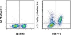
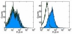
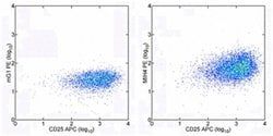
- Applications Reported: The RMP1-30 antibody has been reported for use in flow cytometric analysis
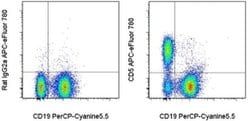
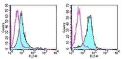
- Applications Tested: This RMP1-30 antibody has been tested by flow cytometric analysis of Con A-stimulated mouse splenocytes
- This can be used at less than or equal to 1 μg per test
- A test is defined as the amount (μg) of antibody that will stain a cell sample in a final volume of 100 μL
- Cell number should be determined empirically but can range from 10^5 to 10^8 cells/test
- It is recommended that the antibody be carefully titrated for optimal performance in the assay of interest
- Excitation: 488-561 nm; Emission: 578 nm; Laser: Blue Laser, Green Laser, Yellow-Green Laser
- Filtration: 0.2 μm post-manufacturing filtered
- Cell-mediated immune responses are initiated by T lymphocytes that are themselves stimulated by cognate peptides bound to MHC molecules on antig en-presenting cells (APC)
- T-cell activation is generally self-limited as activated T cells express receptors such as PD-1 (also known as PDCD-1) that mediate inhibitory signals from the APC
- PD-1 can bind two different but related ligands, PDL-1 and PDL-2
- Upon binding to either of these ligands, signals generated by PD-1 inhibit the activation of the immune response in the absence of "edanger signals"e such as LPS or other molecules associated with bacteria or other pathogens
- Evidence for this is seen in PD1-null mice who exhibit hyperactivated immune systems and autoimmune diseases
- Despite its predicted molecular weight, PD-1 often migrates at higher molecular weight in SDS-PAGE.
Compare Similar Items
Show Difference
Antigen: CD279 (PD-1)
Classification: Monoclonal
Concentration: 0.2 mg/mL
Formulation: PBS with 0.09% sodium azide; pH 7.2
Gene Accession No.: Q02242
Gene Symbols: Pdcd1
Purification Method: Affinity chromatography
Regulatory Status: RUO
Gene ID (Entrez): 18566
Content And Storage: 4° C, store in dark, DO NOT FREEZE!
Form: Liquid
Applications: Flow Cytometry
Clone: RMP1-30
Conjugate: PE
Gene: Pdcd1
Gene Alias: CD279; EGK_05005; hPD1; hPD-1; hPD-l; hSLE1; Ly101; mPD-1; PD1; PD-1; Pdc1; Pdcd1; programmed cell death 1; programmed cell death 1 protein; programmed cell death protein 1; programmed cell death protein 1-like; programmed death 1; Protein PD1; protein PD-1; sCD279; SLEB2; soluble CD279; systemic lupus erythematosus susceptibility 2
Host Species: Rat
Quantity: 100 μg
Primary or Secondary: Primary
Target Species: Mouse
Product Type: Antibody
Isotype: IgG2b κ
Antigen:
CD279 (PD-1)
Classification:
Monoclonal
Concentration:
0.2 mg/mL
Formulation:
PBS with 0.09% sodium azide; pH 7.2
Gene Accession No.:
Q02242
Gene Symbols:
Pdcd1
Purification Method:
Affinity chromatography
Regulatory Status:
RUO
Gene ID (Entrez):
18566
Content And Storage:
4° C, store in dark, DO NOT FREEZE!
Form:
Liquid
Applications:
Flow Cytometry
Clone:
RMP1-30
Conjugate:
PE
Gene:
Pdcd1
Gene Alias:
CD279; EGK_05005; hPD1; hPD-1; hPD-l; hSLE1; Ly101; mPD-1; PD1; PD-1; Pdc1; Pdcd1; programmed cell death 1; programmed cell death 1 protein; programmed cell death protein 1; programmed cell death protein 1-like; programmed death 1; Protein PD1; protein PD-1; sCD279; SLEB2; soluble CD279; systemic lupus erythematosus susceptibility 2
Host Species:
Rat
Quantity:
100 μg
Primary or Secondary:
Primary
Target Species:
Mouse
Product Type:
Antibody
Isotype:
IgG2b κ
Antigen: CD5
Classification: Monoclonal
Concentration: 0.2 mg/mL
Formulation: PBS with 0.09% sodium azide; pH 7.2
Gene Accession No.: P13379
Gene Symbols: Cd5
Purification Method: Affinity chromatography
Regulatory Status: RUO
Gene ID (Entrez): 12507
Content And Storage: 4° C, store in dark, DO NOT FREEZE!
Form: Liquid
Applications: Flow Cytometry
Clone: 53-7.3
Conjugate: APC-eFluor 780
Gene: Cd5
Gene Alias: alanyl (membrane) aminopeptidase; alanyl aminopeptidase; alanyl aminopeptidase, membrane; aminopeptidase M; Aminopeptidase N; ANPEP; AP-M; APN; AP-N; CD antigen CD5; CD13; CD28; CD28 antigen (Tp44); CD28 molecule; Cd5; CD5 antigen; CD5 antigen (p56 62); CD5 antigen (p56-62); CD5 antigen p56-62; CD5 molecule; CD5 protein; CD74 antigen, invariant polypeptide of major; cell surface protein; costimulatory molecule B7 receptor CD28; fCD5; GP150; hAPN; LAP1; LEU1; Ly-1; Ly12; Ly-12; LyA; Ly-A; Lymphocyte antigen 1; Lymphocyte antigen CD5; lymphocyte antigen T1/Leu-1; Lyt-1; membrane alanyl aminopeptidase; microsomal aminopeptidase; Myeloid plasma membrane glycoprotein CD13; P150; Pan T cell; PEPN; T1; T-cell costimulatory molecule CD28; T-cell surface glycoprotein CD5; T-cell-specific surface glycoprotein CD28; unnamed protein product
Host Species: Rat
Quantity: 100 μg
Primary or Secondary: Primary
Target Species: Mouse
Product Type: Antibody
Isotype: IgG2a κ
Antigen:
CD5
Classification:
Monoclonal
Concentration:
0.2 mg/mL
Formulation:
PBS with 0.09% sodium azide; pH 7.2
Gene Accession No.:
P13379
Gene Symbols:
Cd5
Purification Method:
Affinity chromatography
Regulatory Status:
RUO
Gene ID (Entrez):
12507
Content And Storage:
4° C, store in dark, DO NOT FREEZE!
Form:
Liquid
Applications:
Flow Cytometry
Clone:
53-7.3
Conjugate:
APC-eFluor 780
Gene:
Cd5
Gene Alias:
alanyl (membrane) aminopeptidase; alanyl aminopeptidase; alanyl aminopeptidase, membrane; aminopeptidase M; Aminopeptidase N; ANPEP; AP-M; APN; AP-N; CD antigen CD5; CD13; CD28; CD28 antigen (Tp44); CD28 molecule; Cd5; CD5 antigen; CD5 antigen (p56 62); CD5 antigen (p56-62); CD5 antigen p56-62; CD5 molecule; CD5 protein; CD74 antigen, invariant polypeptide of major; cell surface protein; costimulatory molecule B7 receptor CD28; fCD5; GP150; hAPN; LAP1; LEU1; Ly-1; Ly12; Ly-12; LyA; Ly-A; Lymphocyte antigen 1; Lymphocyte antigen CD5; lymphocyte antigen T1/Leu-1; Lyt-1; membrane alanyl aminopeptidase; microsomal aminopeptidase; Myeloid plasma membrane glycoprotein CD13; P150; Pan T cell; PEPN; T1; T-cell costimulatory molecule CD28; T-cell surface glycoprotein CD5; T-cell-specific surface glycoprotein CD28; unnamed protein product
Host Species:
Rat
Quantity:
100 μg
Primary or Secondary:
Primary
Target Species:
Mouse
Product Type:
Antibody
Isotype:
IgG2a κ
Antigen: CD253 (TRAIL)
Classification: Monoclonal
Concentration: 5 μL/Test
Formulation: PBS with 0.2% BSA and 0.09% sodium azide; pH 7.2
Gene Accession No.: P50591
Gene Symbols: TNFSF10
Purification Method: Affinity chromatography
Regulatory Status: RUO
Gene ID (Entrez): 8743
Content And Storage: 4° C, store in dark, DO NOT FREEZE!
Form: Liquid
Applications: Flow Cytometry
Clone: RIK-2
Conjugate: PE
Gene: TNFSF10
Gene Alias: A330042I21Rik; AI448571; Apo-2 ligand; APO2L; Apo-2L; CD253; CD253 antigen; chemokine tumor necrosis factor ligand superfamily member 10; Ly81; Protein TRAIL; TL2; TN; TNF superfamily member 10; TNF-related apoptosis inducing ligand; TNF-related apoptosis inducing ligand TRAIL; TNF-related apoptosis-inducing ligand; Tnfsf10; TNLG6A; TRAIL; TRAIL protein; TRAIL/APO2L; tumor necrosis factor (ligand) family, member 10; tumor necrosis factor (ligand) superfamily member 10; tumor necrosis factor (ligand) superfamily, member 10; tumor necrosis factor apoptosis-inducing ligand splice variant delta; tumor necrosis factor ligand 6A; tumor necrosis factor ligand superfamily member 10; tumor necrosis factor superfamily member 10
Host Species: Mouse
Quantity: 100 Tests
Primary or Secondary: Primary
Target Species: Human
Product Type: Antibody
Isotype: IgG1 κ
Antigen:
CD253 (TRAIL)
Classification:
Monoclonal
Concentration:
5 μL/Test
Formulation:
PBS with 0.2% BSA and 0.09% sodium azide; pH 7.2
Gene Accession No.:
P50591
Gene Symbols:
TNFSF10
Purification Method:
Affinity chromatography
Regulatory Status:
RUO
Gene ID (Entrez):
8743
Content And Storage:
4° C, store in dark, DO NOT FREEZE!
Form:
Liquid
Applications:
Flow Cytometry
Clone:
RIK-2
Conjugate:
PE
Gene:
TNFSF10
Gene Alias:
A330042I21Rik; AI448571; Apo-2 ligand; APO2L; Apo-2L; CD253; CD253 antigen; chemokine tumor necrosis factor ligand superfamily member 10; Ly81; Protein TRAIL; TL2; TN; TNF superfamily member 10; TNF-related apoptosis inducing ligand; TNF-related apoptosis inducing ligand TRAIL; TNF-related apoptosis-inducing ligand; Tnfsf10; TNLG6A; TRAIL; TRAIL protein; TRAIL/APO2L; tumor necrosis factor (ligand) family, member 10; tumor necrosis factor (ligand) superfamily member 10; tumor necrosis factor (ligand) superfamily, member 10; tumor necrosis factor apoptosis-inducing ligand splice variant delta; tumor necrosis factor ligand 6A; tumor necrosis factor ligand superfamily member 10; tumor necrosis factor superfamily member 10
Host Species:
Mouse
Quantity:
100 Tests
Primary or Secondary:
Primary
Target Species:
Human
Product Type:
Antibody
Isotype:
IgG1 κ

Antigen: __
Classification: __
Concentration: __
Formulation: __
Gene Accession No.: __
Gene Symbols: __
Purification Method: __
Regulatory Status: __
Gene ID (Entrez): __
Content And Storage: __
Form: __
Applications: __
Clone: __
Conjugate: __
Gene: __
Gene Alias: __
Host Species: __
Quantity: __
Primary or Secondary: __
Target Species: __
Product Type: __
Isotype: __
Antigen:
__
Classification:
__
Concentration:
__
Formulation:
__
Gene Accession No.:
__
Gene Symbols:
__
Purification Method:
__
Regulatory Status:
__
Gene ID (Entrez):
__
Content And Storage:
__
Form:
__
Applications:
__
Clone:
__
Conjugate:
__
Gene:
__
Gene Alias:
__
Host Species:
__
Quantity:
__
Primary or Secondary:
__
Target Species:
__
Product Type:
__
Isotype:
__