50-223-8246
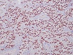
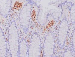
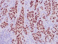
SALL4 Recombinant Rabbit Monoclonal Antibody (ZR276), RAbMono™, Zeta
Manufacturer: ZETA CORPORATION
Select a Size
| Pack Size | SKU | Availability | Price |
|---|---|---|---|
| Each of 1 | 50-223-8246-Each-of-1 | In Stock | ₹ 7,476.00 |
50-223-8246 - Each of 1
In Stock
Quantity
1
Base Price: ₹ 7,476.00
GST (18%): ₹ 1,345.68
Total Price: ₹ 8,821.68
Antigen
SALL4
Classification
Recombinant Monoclonal
Concentration
100 μg/mL
Formulation
tris with BSA, NP-40 and <0.1% sodium azide
Gene Accession No.
Q9UJQ4
Gene Symbols
SALL4
Immunogen
Recombinant fragment (around aa1-185) of human MIC2 protein
Quantity
100 μL
Primary or Secondary
Primary
Target Species
Human
Product Type
Antibody
Isotype
IgG
Applications
Immunohistochemistry (Paraffin)
Clone
ZR276
Conjugate
Unconjugated
Gene
SALL4
Gene Alias
5730441M18Rik; AA407717; AL022809; AW536104; C330011P20Rik; C78083; C78563; dJ1112F19.1; DRRS; HSAL4; SALL4; sal-like 4; sal-like 4 (Drosophila); sal-like protein 4; spalt like transcription factor 4; spalt-like transcription factor 4; Tex20; Zinc finger protein 797; zinc finger protein SALL4; ZNF797
Host Species
Rabbit
Purification Method
Protein A
Regulatory Status
RUO
Gene ID (Entrez)
57167
Content And Storage
2-8°C
Form
Liquid
Description
- This product is diluted and in a ready-to-use formulation
- A recommended positive control tissue for this product is Seminoma, however positive controls are not limited to this tissue type
- The primary antibody is intended for laboratory professional use in the detection of the corresponding protein in formalin-fixed, paraffin-embedded tissue stained in manual qualitative immunohistochemistry (IHC) testing
- This antibody is intended to be used after the primary diagnosis of tumor has been made by conventional histopathology using non-immunological histochemical stains
- Antibody is used with formalin-fixed and paraffin-embedded sections
- Pretreatment of deparaffinized tissue with heat-induced epitope retrieval or enzymatic retrieval is recommended
- In general, immunohistochemical (IHC) staining techniques allow for the visualization of antigens via the sequential application of a specific antibody to the antigen (primary antibody), a secondary antibody to the primary antibody (link antibody), an enzyme complex and a chromogenic substrate with interposed washing steps
- The enzymatic activation of the chromogen results in a visible reaction product at the antigen site
- Results are interpreted using a light microscope and aid in the differential diagnosis of pathophysiological processes, which may or may not be associated with a particular antigen
- A positive tissue control must be run with every staining procedure performed
- This tissue may contain both positive and negative staining cells or tissue components and serve as both the positive and negative control tissue
- External Positive control materials should be fresh autopsy/biopsy/surgical specimens fixed, processed and embedded as soon as possible in the same manner as the patient sample (s)
- Positive tissue controls are indicative of correctly prepared tissues and proper staining methods
- The tissues used for the external positive control materials should be selected from the patient specimens with well-characterized low levels of the positive target activity that gives weak positive staining
- The low level of positivity for external positive controls is designed to ensure detection of subtle changes in the primary antibody sensitivity from instability or problems with the staining methodology
- A tissue with weak positive staining is more suitable for optimal quality control and for detecting minor levels of reagent degradation
- Internal or external negative control tissue may be used depending on the guidelines and policies that govern the organization to which the end user belongs to
- The variety of cell types present in many tissue sections offers internal negative control sites, but this should be verified by the user
- The components that do not stain should demonstrate the absence of specific staining, and provide an indication of non-specific background staining
- If specific staining occurs in the negative tissue control sites, results with the patient specimens must be considered invalid.
Compare Similar Items
Show Difference
Antigen: SALL4
Classification: Recombinant Monoclonal
Concentration: 100 μg/mL
Formulation: tris with BSA, NP-40 and <0.1% sodium azide
Gene Accession No.: Q9UJQ4
Gene Symbols: SALL4
Immunogen: Recombinant fragment (around aa1-185) of human MIC2 protein
Quantity: 100 μL
Primary or Secondary: Primary
Target Species: Human
Product Type: Antibody
Isotype: IgG
Applications: Immunohistochemistry (Paraffin)
Clone: ZR276
Conjugate: Unconjugated
Gene: SALL4
Gene Alias: 5730441M18Rik; AA407717; AL022809; AW536104; C330011P20Rik; C78083; C78563; dJ1112F19.1; DRRS; HSAL4; SALL4; sal-like 4; sal-like 4 (Drosophila); sal-like protein 4; spalt like transcription factor 4; spalt-like transcription factor 4; Tex20; Zinc finger protein 797; zinc finger protein SALL4; ZNF797
Host Species: Rabbit
Purification Method: Protein A
Regulatory Status: RUO
Gene ID (Entrez): 57167
Content And Storage: 2-8°C
Form: Liquid
Antigen:
SALL4
Classification:
Recombinant Monoclonal
Concentration:
100 μg/mL
Formulation:
tris with BSA, NP-40 and <0.1% sodium azide
Gene Accession No.:
Q9UJQ4
Gene Symbols:
SALL4
Immunogen:
Recombinant fragment (around aa1-185) of human MIC2 protein
Quantity:
100 μL
Primary or Secondary:
Primary
Target Species:
Human
Product Type:
Antibody
Isotype:
IgG
Applications:
Immunohistochemistry (Paraffin)
Clone:
ZR276
Conjugate:
Unconjugated
Gene:
SALL4
Gene Alias:
5730441M18Rik; AA407717; AL022809; AW536104; C330011P20Rik; C78083; C78563; dJ1112F19.1; DRRS; HSAL4; SALL4; sal-like 4; sal-like 4 (Drosophila); sal-like protein 4; spalt like transcription factor 4; spalt-like transcription factor 4; Tex20; Zinc finger protein 797; zinc finger protein SALL4; ZNF797
Host Species:
Rabbit
Purification Method:
Protein A
Regulatory Status:
RUO
Gene ID (Entrez):
57167
Content And Storage:
2-8°C
Form:
Liquid
Antigen: CD68
Classification: Recombinant Monoclonal
Concentration: 1 μg/mL
Formulation: tris with BSA, NP-40 and <0.1% sodium azide
Gene Accession No.: P34810
Gene Symbols: Cd68
Immunogen: Recombinant fragment (around aa1-200) of human CD68
Quantity: 7 mL
Primary or Secondary: Primary
Target Species: Human
Product Type: Antibody
Isotype: IgG
Applications: Immunohistochemistry (Paraffin)
Clone: ZR302
Conjugate: Unconjugated
Gene: Cd68
Gene Alias: CD68; CD68 antigen; CD68 molecule; GP110; Lamp4; macrophage antigen CD68; macrosialin; Scard1; scavenger receptor class D, member 1; similar to CD68 antigen
Host Species: Rabbit
Purification Method: Protein A
Regulatory Status: RUO
Gene ID (Entrez): 968
Content And Storage: 2-8°C
Form: Liquid
Antigen:
CD68
Classification:
Recombinant Monoclonal
Concentration:
1 μg/mL
Formulation:
tris with BSA, NP-40 and <0.1% sodium azide
Gene Accession No.:
P34810
Gene Symbols:
Cd68
Immunogen:
Recombinant fragment (around aa1-200) of human CD68
Quantity:
7 mL
Primary or Secondary:
Primary
Target Species:
Human
Product Type:
Antibody
Isotype:
IgG
Applications:
Immunohistochemistry (Paraffin)
Clone:
ZR302
Conjugate:
Unconjugated
Gene:
Cd68
Gene Alias:
CD68; CD68 antigen; CD68 molecule; GP110; Lamp4; macrophage antigen CD68; macrosialin; Scard1; scavenger receptor class D, member 1; similar to CD68 antigen
Host Species:
Rabbit
Purification Method:
Protein A
Regulatory Status:
RUO
Gene ID (Entrez):
968
Content And Storage:
2-8°C
Form:
Liquid
Antigen: GATA3
Classification: Recombinant Monoclonal
Concentration: 200 μg/mL
Formulation: tris with BSA, NP-40 and <0.1% sodium azide
Gene Accession No.: P23771
Gene Symbols: GATA3
Immunogen: Recombinant full-length human GATA-3 protein
Quantity: 100 μL
Primary or Secondary: Primary
Target Species: Human
Product Type: Antibody
Isotype: IgG
Applications: Immunohistochemistry (Paraffin)
Clone: ZR358
Conjugate: Unconjugated
Gene: GATA3
Gene Alias: EGK_19445; GATA binding factor-3; GATA binding protein 3; GATA binding protein-3; GATA3; Gata-3; GATA-binding factor 3; GATA-binding protein 3; HDR; HDRS; jal; MGC2346; MGC5199; MGC5445; Trans-acting T-cell-specific transcription factor GATA-3; Transacting T-cell-specific transcription factor GATA-3
Host Species: Rabbit
Purification Method: Protein A
Regulatory Status: RUO
Gene ID (Entrez): 2625
Content And Storage: 2-8°C
Form: Liquid
Antigen:
GATA3
Classification:
Recombinant Monoclonal
Concentration:
200 μg/mL
Formulation:
tris with BSA, NP-40 and <0.1% sodium azide
Gene Accession No.:
P23771
Gene Symbols:
GATA3
Immunogen:
Recombinant full-length human GATA-3 protein
Quantity:
100 μL
Primary or Secondary:
Primary
Target Species:
Human
Product Type:
Antibody
Isotype:
IgG
Applications:
Immunohistochemistry (Paraffin)
Clone:
ZR358
Conjugate:
Unconjugated
Gene:
GATA3
Gene Alias:
EGK_19445; GATA binding factor-3; GATA binding protein 3; GATA binding protein-3; GATA3; Gata-3; GATA-binding factor 3; GATA-binding protein 3; HDR; HDRS; jal; MGC2346; MGC5199; MGC5445; Trans-acting T-cell-specific transcription factor GATA-3; Transacting T-cell-specific transcription factor GATA-3
Host Species:
Rabbit
Purification Method:
Protein A
Regulatory Status:
RUO
Gene ID (Entrez):
2625
Content And Storage:
2-8°C
Form:
Liquid

Antigen: SCN1B
Classification: Polyclonal
Concentration: 267 μg/mL
Formulation: __
Gene Accession No.: __
Gene Symbols: __
Immunogen: __
Quantity: 150 μL
Primary or Secondary: Primary
Target Species: Human, Rat, Mouse
Product Type: __
Isotype: IgG
Applications: Western Blot, Immunohistochemistry, ELISA
Clone: __
Conjugate: Unconjugated
Gene: __
Gene Alias: __
Host Species: Rabbit
Purification Method: __
Regulatory Status: __
Gene ID (Entrez): 6324
Content And Storage: __
Form: __
Antigen:
SCN1B
Classification:
Polyclonal
Concentration:
267 μg/mL
Formulation:
__
Gene Accession No.:
__
Gene Symbols:
__
Immunogen:
__
Quantity:
150 μL
Primary or Secondary:
Primary
Target Species:
Human, Rat, Mouse
Product Type:
__
Isotype:
IgG
Applications:
Western Blot, Immunohistochemistry, ELISA
Clone:
__
Conjugate:
Unconjugated
Gene:
__
Gene Alias:
__
Host Species:
Rabbit
Purification Method:
__
Regulatory Status:
__
Gene ID (Entrez):
6324
Content And Storage:
__
Form:
__