Cytochrome c Antibody (CTC05), Novus Biologicals™
Mouse Monoclonal Antibody
Manufacturer: Fischer Scientific
The price for this product is unavailable. Please request a quote
Antigen
Cytochrome c
Concentration
0.2mg/mL
Applications
Western Blot, Flow Cytometry, Immunohistochemistry (Paraffin), Immunofluorescence
Conjugate
Unconjugated
Host Species
Mouse
Research Discipline
Apoptosis, Cellular Markers, Cholesterol Metabolism, Core ESC Like Genes, Lipid and Metabolism, Mitochondrial Markers, Stem Cell Markers
Formulation
1.0mM PBS and 0.05% BSA with 0.05% Sodium Azide
Gene ID (Entrez)
54205
Immunogen
Recombinant cytochrome c protein
Primary or Secondary
Primary
Content And Storage
Store at 4C.
Molecular Weight of Antigen
15 kDa
Clone
CTC05
Dilution
Western Blot 0.5 - 1.0 ug/ml, Flow Cytometry 0.5 - 1 ug/million cells in 0.1 ml, Immunohistochemistry-Paraffin 0.25 - 0.5 ug/ml, Immunofluorescence 0.5 - 1.0 ug/ml
Classification
Monoclonal
Form
Purified
Regulatory Status
RUO
Target Species
Human, Mouse, Rat, Amphibian, Avian, Canine, Drosophila, Equine
Gene Alias
CYCHCS, cytochrome c, cytochrome c, somatic, THC4
Gene Symbols
CYCS
Isotype
IgG2b κ
Purification Method
Protein A or G purified
Test Specificity
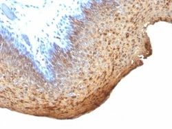
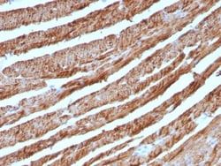
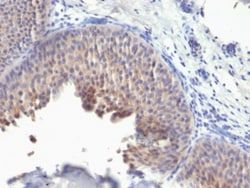
Cytochrome C is a well-characterized mobile electron transport protein that is essential to energy conversion in all aerobic organisms. In mammalian cells, this highly conserved protein is normally localized to the mitochondrial inter-membrane space. More recent studies have identified cytosolic cytochrome c as a factor necessary for activation of apoptosis. During apoptosis, cytochrome c is trans-located from the mitochondrial membrane to the cytosol, where it is required for activation of caspase-3 (CPP32). Overexpression of Bcl-2 has been shown to prevent the translocation of cytochrome c, thereby blocking the apoptotic process. Overexpression of Bax has been shown to induce the release of cytochrome c and to induce cell death. The release of cytochrome c from the mitochondria is thought to trigger an apoptotic cascade, whereby Apaf-1 binds to Apaf-3 (caspase-9) in a cytochrome c-dependent manner, leading to caspase-9 cleavage of caspase-3.
Description
- Ensure accurate, reproducible results in Western Blot, Flow Cytometry, Immunohistochemistry (Paraffin), Immunofluorescence Cytochrome c Monoclonal specifically detects Cytochrome c in Human, Mouse, Rat, Amphibian, Avian, Canine, Drosophila, Equine, Pigeon samples
- It is validated for Western Blot, Flow Cytometry, Immunohistochemistry, Immunocytochemistry/Immunofluorescence, Immunohistochemistry-Paraffin.
Compare Similar Items
Show Difference
Antigen: Cytochrome c
Concentration: 0.2mg/mL
Applications: Western Blot, Flow Cytometry, Immunohistochemistry (Paraffin), Immunofluorescence
Conjugate: Unconjugated
Host Species: Mouse
Research Discipline: Apoptosis, Cellular Markers, Cholesterol Metabolism, Core ESC Like Genes, Lipid and Metabolism, Mitochondrial Markers, Stem Cell Markers
Formulation: 1.0mM PBS and 0.05% BSA with 0.05% Sodium Azide
Gene ID (Entrez): 54205
Immunogen: Recombinant cytochrome c protein
Primary or Secondary: Primary
Content And Storage: Store at 4C.
Molecular Weight of Antigen: 15 kDa
Clone: CTC05
Dilution: Western Blot 0.5 - 1.0 ug/ml, Flow Cytometry 0.5 - 1 ug/million cells in 0.1 ml, Immunohistochemistry-Paraffin 0.25 - 0.5 ug/ml, Immunofluorescence 0.5 - 1.0 ug/ml
Classification: Monoclonal
Form: Purified
Regulatory Status: RUO
Target Species: Human, Mouse, Rat, Amphibian, Avian, Canine, Drosophila, Equine
Gene Alias: CYCHCS, cytochrome c, cytochrome c, somatic, THC4
Gene Symbols: CYCS
Isotype: IgG2b κ
Purification Method: Protein A or G purified
Test Specificity: Cytochrome C is a well-characterized mobile electron transport protein that is essential to energy conversion in all aerobic organisms. In mammalian cells, this highly conserved protein is normally localized to the mitochondrial inter-membrane space. More recent studies have identified cytosolic cytochrome c as a factor necessary for activation of apoptosis. During apoptosis, cytochrome c is trans-located from the mitochondrial membrane to the cytosol, where it is required for activation of caspase-3 (CPP32). Overexpression of Bcl-2 has been shown to prevent the translocation of cytochrome c, thereby blocking the apoptotic process. Overexpression of Bax has been shown to induce the release of cytochrome c and to induce cell death. The release of cytochrome c from the mitochondria is thought to trigger an apoptotic cascade, whereby Apaf-1 binds to Apaf-3 (caspase-9) in a cytochrome c-dependent manner, leading to caspase-9 cleavage of caspase-3.
Antigen: Cytokeratin 10
Concentration: 0.2mg/mL
Applications: Flow Cytometry, Immunohistochemistry (Paraffin), Immunofluorescence
Conjugate: Unconjugated
Host Species: Mouse
Research Discipline: Cytoskeleton Markers
Formulation: 10mM PBS and 0.05% BSA with 0.05% Sodium Azide
Gene ID (Entrez): 3858
Immunogen: Recombinant human KRT10 protein
Primary or Secondary: Primary
Content And Storage: Store at 4C.
Molecular Weight of Antigen: 56.5 kDa
Clone: KRT10/1275
Dilution: Flow Cytometry 0.5 - 1 ug/million cells in 0.1 ml, Immunohistochemistry-Paraffin 0.5 - 1.0 ug/ml, Immunofluorescence 0.5 - 1.0 ug/ml
Classification: Monoclonal
Form: Purified
Regulatory Status: RUO
Target Species: Human
Gene Alias: BCIE, BIE, CK10, CK-10, cytokeratin 10, Cytokeratin-10, EHK, K10keratosis palmaris et plantaris, keratin 10, Keratin 10 (epidermolytic hyperkeratosis; keratosis palmaris et plantaris), keratin, type I cytoskeletal 10, keratin-10, KPP
Gene Symbols: KRT10
Isotype: IgG1 κ
Purification Method: Protein A or G purified
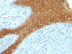
Test Specificity: This MAb recognizes a protein of 56.5kDa, identified as cytokeratin 10 (CK10). CK10 is expressed in all suprabasal layers of the epidermis. In the epidermis, expression of CK10 strictly parallels the extent of differentiation; it is absent in the basal layer, appears in the first suprabasal layers and increases in concentration towards the granular layer. However, CK10 is rarely detected in early stages of vulvar squamous carcinomas (tumors less than 2 cm, clinical stage I) regardless of the tumor grade. In larger and more advanced tumors (greater than 2 cm, clinical stages II and III), CK10 is detected very frequently. Expression of CK10 is related to maturation of malignant keratinocytes, being preferentially detected in more-differentiated parts.
Antigen: Cytokeratin 10
Concentration: 0.2 mg/mL
Applications: Flow Cytometry, Immunohistochemistry (Paraffin), Immunofluorescence
Conjugate: Unconjugated
Host Species: Mouse
Research Discipline: Cytoskeleton Markers
Formulation: 10mM PBS and 0.05% BSA with 0.05% Sodium Azide
Gene ID (Entrez): 3858
Immunogen: Recombinant human KRT10 protein
Primary or Secondary: Primary
Content And Storage: Store at 4C.
Molecular Weight of Antigen: 56.5 kDa
Clone: KRT10/844 + KRT10/1275
Dilution: Flow Cytometry 0.5 - 1 ug/million cells in 0.1 ml, Immunohistochemistry-Paraffin 0.5 - 1.0 ug/ml, Immunofluorescence 0.5 - 1.0 ug/ml
Classification: Monoclonal
Form: Purified
Regulatory Status: RUO
Target Species: Human
Gene Alias: BCIE, BIE, CK10, CK-10, cytokeratin 10, Cytokeratin-10, EHK, K10keratosis palmaris et plantaris, keratin 10, Keratin 10 (epidermolytic hyperkeratosis; keratosis palmaris et plantaris), keratin, type I cytoskeletal 10, keratin-10, KPP
Gene Symbols: KRT10
Isotype: IgG1 κ
Purification Method: Protein A or G purified
Test Specificity: This MAb recognizes a protein of 56.5kDa, identified as cytokeratin 10 (CK10). CK10 is expressed in all suprabasal layers of the epidermis. In the epidermis, expression of CK10 strictly parallels the extent of differentiation; it is absent in the basal layer, appears in the first suprabasal layers and increases in concentration towards the granular layer. However, CK10 is rarely detected in early stages of vulvar squamous carcinomas (tumors less than 2 cm, clinical stage I) regardless of the tumor grade. In larger and more advanced tumors (greater than 2 cm, clinical stages II and III), CK10 is detected very frequently. Expression of CK10 is related to maturation of malignant keratinocytes, being preferentially detected in more-differentiated parts.