Cytokeratin 10/13 Antibody (SPM262), Novus Biologicals™
Mouse Monoclonal Antibody
Manufacturer: Fischer Scientific
The price for this product is unavailable. Please request a quote
Antigen
Cytokeratin 10/13
Concentration
0.2mg/mL
Applications
Western Blot, Flow Cytometry, Immunohistochemistry (Paraffin), Immunofluorescence
Conjugate
Unconjugated
Host Species
Mouse
Research Discipline
Cytoskeleton Markers
Formulation
10mM PBS and 0.05% BSA with 0.05% Sodium Azide
Gene ID (Entrez)
3858
Immunogen
Cytoskeletal preparation extracted from human ectocervical epithelium.
Primary or Secondary
Primary
Content And Storage
Store at 4C.
Clone
SPM262
Dilution
Western Blot 0.25 - 0.5 ug/ml, Flow Cytometry 0.5 - 1 ug/million cells in 0.1 ml, Immunohistochemistry-Paraffin 0.5 - 1.0 ug/ml, Immunofluorescence 0.5 - 1.0 ug/ml
Classification
Monoclonal
Form
Purified
Regulatory Status
RUO
Target Species
Human, Feline
Gene Alias
BCIE, BIE, CK10, CK-10, cytokeratin 10, Cytokeratin-10, EHK, K10keratosis palmaris et plantaris, keratin 10, keratin, type I cytoskeletal 10, keratin-10, KPP
Gene Symbols
KRT10
Isotype
IgG2a κ
Purification Method
Protein A or G purified
Test Specificity
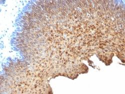
This antibody recognizes cytokeratin 10 (56.5kDa) and cytokeratin 13 (53kDa) in Western blotting. It recognizes only cytokeratin 13 in formalin-fixed, paraffin-embedded tissue sections. It does not react with cytokeratin 10 positive, cytokeratin 13 negative epithelia such as epidermis. However, on frozen sections this MAb serves as differentiation-related marker of all stratified epithelia; it stains all suprabasal cells in both cornifying and non-cornifying stratified epithelia and more differentiated cells of squamous carcinomas.
Description
- Ensure accurate, reproducible results in Western Blot, Flow Cytometry, Immunohistochemistry (Paraffin), Immunofluorescence Cytokeratin 10/13 Monoclonal specifically detects Cytokeratin 10/13 in Human, Feline samples
- It is validated for Western Blot, Flow Cytometry, Immunohistochemistry, Immunocytochemistry/Immunofluorescence, Immunohistochemistry-Paraffin, Immunofluorescence.
Compare Similar Items
Show Difference
Antigen: Cytokeratin 10/13
Concentration: 0.2mg/mL
Applications: Western Blot, Flow Cytometry, Immunohistochemistry (Paraffin), Immunofluorescence
Conjugate: Unconjugated
Host Species: Mouse
Research Discipline: Cytoskeleton Markers
Formulation: 10mM PBS and 0.05% BSA with 0.05% Sodium Azide
Gene ID (Entrez): 3858
Immunogen: Cytoskeletal preparation extracted from human ectocervical epithelium.
Primary or Secondary: Primary
Content And Storage: Store at 4C.
Clone: SPM262
Dilution: Western Blot 0.25 - 0.5 ug/ml, Flow Cytometry 0.5 - 1 ug/million cells in 0.1 ml, Immunohistochemistry-Paraffin 0.5 - 1.0 ug/ml, Immunofluorescence 0.5 - 1.0 ug/ml
Classification: Monoclonal
Form: Purified
Regulatory Status: RUO
Target Species: Human, Feline
Gene Alias: BCIE, BIE, CK10, CK-10, cytokeratin 10, Cytokeratin-10, EHK, K10keratosis palmaris et plantaris, keratin 10, keratin, type I cytoskeletal 10, keratin-10, KPP
Gene Symbols: KRT10
Isotype: IgG2a κ
Purification Method: Protein A or G purified
Test Specificity: This antibody recognizes cytokeratin 10 (56.5kDa) and cytokeratin 13 (53kDa) in Western blotting. It recognizes only cytokeratin 13 in formalin-fixed, paraffin-embedded tissue sections. It does not react with cytokeratin 10 positive, cytokeratin 13 negative epithelia such as epidermis. However, on frozen sections this MAb serves as differentiation-related marker of all stratified epithelia; it stains all suprabasal cells in both cornifying and non-cornifying stratified epithelia and more differentiated cells of squamous carcinomas.
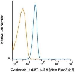
Antigen: Cytokeratin 14
Concentration: 0.2mg/mL
Applications: Flow Cytometry, Immunohistochemistry (Paraffin), Immunofluorescence
Conjugate: Unconjugated
Host Species: Mouse
Research Discipline: Cytoskeleton Markers
Formulation: 10mM PBS and 0.05% BSA with 0.05% Sodium Azide
Gene ID (Entrez): 3861
Immunogen: Recombinant full-length human KRT14 protein
Primary or Secondary: Primary
Content And Storage: Store at 4C.
Clone: KRT14/532
Dilution: Flow Cytometry 0.5 - 1 ug/million cells in 0.1 ml, Immunohistochemistry-Paraffin 0.5 - 1.0 ug/ml, Immunofluorescence 0.5 - 1.0 ug/ml
Classification: Monoclonal
Form: Purified
Regulatory Status: RUO
Target Species: Human, Mouse, Rat
Gene Alias: CK14, CK-14, cytokeratin 14, cytokeratin-14, EBS3, EBS4, K14, keratin 14, keratin 14 (epidermolysis bullosa simplex, Dowling-Meara, Koebner), keratin, type I cytoskeletal 14, Keratin-14, NFJ
Gene Symbols: KRT14
Isotype: IgG3
Purification Method: Protein A or G purified
Test Specificity: Cytokeratin 14 (CK14) belongs to the type I (or A or acidic) subfamily of low molecular weight keratins and exists in combination with keratin 5 (type II or B or basic). CK14 is found in basal cells of squamous epithelia, some glandular epithelia, myoepithelium, and mesothelial cells. Anti-CK14 is useful in differentiating squamous cell carcinomas from poorly differentiated epithelial tumors. Anti-CK14 is one of the specific basal markers for distinguishing between basal and non-basal subtypes of breast carcinomas. Anti-CK14 is also a good marker for differentiation of intraductal from invasive salivary duct carcinoma by the positive staining of basal cells surrounding the in-situ neoplasm as well as for differentiation of benign prostate from prostate carcinoma. Furthermore, this antibody has been useful in separating oncocytic tumors of the kidney from its renal mimics, and in identifying metaplastic carcinomas of the breast.
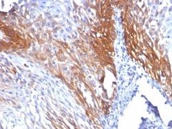
Antigen: Cytokeratin 17
Concentration: 0.2mg/mL
Applications: Flow Cytometry, Immunohistochemistry (Paraffin), SDS-Page, Immunofluorescence
Conjugate: Unconjugated
Host Species: Mouse
Research Discipline: Cancer
Formulation: 10mM PBS and 0.05% BSA with 0.05% Sodium Azide
Gene ID (Entrez): 3872
Immunogen: Recombinant full-length human KRT17 protein
Primary or Secondary: Primary
Content And Storage: Store at 4C.
Clone: KRT17/778
Dilution: Flow Cytometry 0.5 - 1 ug/million cells in 0.1 ml, Immunohistochemistry-Paraffin 0.5 - 1.0 ug/ml, SDS-Page, Immunofluorescence 1 - 2 ug/ml
Classification: Monoclonal
Form: Purified
Regulatory Status: RUO
Target Species: Human, Rat, Porcine, Goat, Primate
Gene Alias: 39.1, CK-17, cytokeratin-17, K17, keratin 17, keratin, type I cytoskeletal 17, keratin-17, PC, PC2, PCHC1
Gene Symbols: KRT17
Isotype: IgG2b κ
Purification Method: Protein A or G purified
Test Specificity: Cytokeratin 17 (CK17) is normally expressed in the basal cells of complex epithelia but not in stratified or simple epithelia. Antibody to CK17 is an excellent tool to distinguish myoepithelial cells from luminal epithelium of various glands such as mammary, sweat and salivary. CK17 is expressed in epithelial cells of various origins, such as bronchial epithelial cells and skin appendages. It may be considered as epithelial stem cell marker because CK17 Ab marks basal cell differentiation. CK17 is expressed in SCLC much higher than in LADC. Eighty-five percent of the triple negative breast carcinomas immunoreact with basal cytokeratins including anti-CK17. Also important is that cases of triple negative breast carcinoma with expression of CK17 show an aggressive clinical course. The histologic differentiation of ampullary cancer, intestinal vs. pancreatobiliary, is very important for treatment. Usually anti-CK17 and anti-MUC1 immunoreactivity represents pancreatobiliary subtype whereas