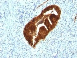
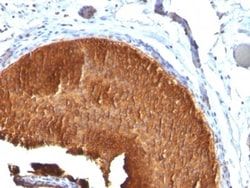
Cytokeratin 18 Antibody (KRT18/836), Novus Biologicals™
Mouse Monoclonal Antibody
Manufacturer: Fischer Scientific
The price for this product is unavailable. Please request a quote
Antigen
Cytokeratin 18
Concentration
0.2mg/mL
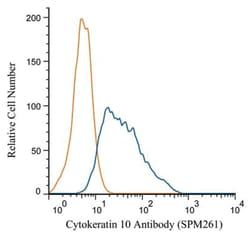
Applications
Flow Cytometry, Immunohistochemistry (Paraffin), Immunofluorescence
Conjugate
Unconjugated
Host Species
Mouse
Research Discipline
Cancer, Cell Biology, Cellular Markers, Cytoskeleton Markers
Formulation
10mM PBS and 0.05% BSA with 0.05% Sodium Azide
Gene ID (Entrez)
3875
Immunogen
Recombinant human KRT18 protein
Primary or Secondary
Primary
Content And Storage
Store at 4C.
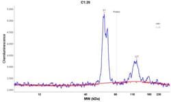
Molecular Weight of Antigen
45 kDa
Clone
KRT18/836
Dilution
Flow Cytometry 0.5 - 1 ug/million cells in 0.1 ml, Immunohistochemistry-Paraffin 2 - 4 ug/ml, Immunofluorescence 1 - 2 ug/ml
Classification
Monoclonal
Form
Purified
Regulatory Status
RUO
Target Species
Human, Canine, Feline, Rat (Negative)
Gene Alias
Cell proliferation-inducing gene 46 protein, cell proliferation-inducing protein 46, CK-18, CYK18, cytokeratin 18, cytokeratin-18, K18, keratin 18, keratin, type I cytoskeletal 18, keratin-18
Gene Symbols
KRT18
Isotype
IgG1
Purification Method
Protein A or G purified
Test Specificity
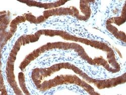
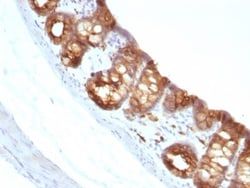
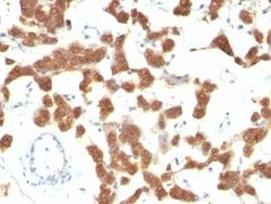
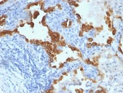
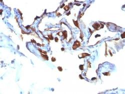
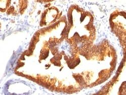
This MAb reacts with a wide variety of simple epithelia. It does not react with stratified squamous epithelia. It reacts with epithelial tumors of the gastrointestinal tract, lung, breast, pancreas, ovary, and thyroid. Cytokeratin 18, which belongs to the type A (acidic) subfamily of low molecular weight keratins, exists in combination with cytokeratin 8. It is reported that tissues from gastrointestinal tract are positive for both cytokeratin 8 and 18 but do not contain cytokeratin 14. Tissues from gastrointestinal tract, respiratory tract and urogenital tract, as well as endocrine and exocrine tissues and mesothelial cells are positive for cytokeratin 18.
Related Products
Description
- Ensure accurate, reproducible results in Flow Cytometry, Immunohistochemistry (Paraffin), Immunofluorescence Cytokeratin 18 Monoclonal specifically detects Cytokeratin 18 in Human, Canine, Feline, Rat (Negative) samples
- It is validated for Western Blot, Flow Cytometry, Immunohistochemistry, Immunocytochemistry/Immunofluorescence, Immunohistochemistry-Paraffin, Protein Array.
Compare Similar Items
Show Difference
Antigen: Cytokeratin 18
Concentration: 0.2mg/mL
Applications: Flow Cytometry, Immunohistochemistry (Paraffin), Immunofluorescence
Conjugate: Unconjugated
Host Species: Mouse
Research Discipline: Cancer, Cell Biology, Cellular Markers, Cytoskeleton Markers
Formulation: 10mM PBS and 0.05% BSA with 0.05% Sodium Azide
Gene ID (Entrez): 3875
Immunogen: Recombinant human KRT18 protein
Primary or Secondary: Primary
Content And Storage: Store at 4C.
Molecular Weight of Antigen: 45 kDa
Clone: KRT18/836
Dilution: Flow Cytometry 0.5 - 1 ug/million cells in 0.1 ml, Immunohistochemistry-Paraffin 2 - 4 ug/ml, Immunofluorescence 1 - 2 ug/ml
Classification: Monoclonal
Form: Purified
Regulatory Status: RUO
Target Species: Human, Canine, Feline, Rat (Negative)
Gene Alias: Cell proliferation-inducing gene 46 protein, cell proliferation-inducing protein 46, CK-18, CYK18, cytokeratin 18, cytokeratin-18, K18, keratin 18, keratin, type I cytoskeletal 18, keratin-18
Gene Symbols: KRT18
Isotype: IgG1
Purification Method: Protein A or G purified
Test Specificity: This MAb reacts with a wide variety of simple epithelia. It does not react with stratified squamous epithelia. It reacts with epithelial tumors of the gastrointestinal tract, lung, breast, pancreas, ovary, and thyroid. Cytokeratin 18, which belongs to the type A (acidic) subfamily of low molecular weight keratins, exists in combination with cytokeratin 8. It is reported that tissues from gastrointestinal tract are positive for both cytokeratin 8 and 18 but do not contain cytokeratin 14. Tissues from gastrointestinal tract, respiratory tract and urogenital tract, as well as endocrine and exocrine tissues and mesothelial cells are positive for cytokeratin 18.
Antigen: Cytokeratin 19
Concentration: 0.2mg/mL
Applications: Flow Cytometry, Immunohistochemistry (Paraffin), Immunofluorescence
Conjugate: Unconjugated
Host Species: Mouse
Research Discipline: Cancer, Cell Biology, Cellular Markers, Cytoskeleton Markers, Neuroscience, Stem Cell Markers
Formulation: 10mM PBS and 0.05% BSA with 0.05% Sodium Azide
Gene ID (Entrez): 3880
Immunogen: Recombinant full-length human KRT19 protein
Primary or Secondary: Primary
Content And Storage: Store at 4C.
Molecular Weight of Antigen: 40 kDa
Clone: KRT19/799 + KRT19/800
Dilution: Flow Cytometry 0.5 - 1 ug/million cells in 0.1 ml, Immunohistochemistry-Paraffin 0.5 - 1.0 ug/ml, Immunofluorescence 1 - 2 ug/ml
Classification: Monoclonal
Form: Purified
Regulatory Status: RUO
Target Species: Human, Mouse
Gene Alias: CK19, CK-19, cytokeratin 19, Cytokeratin-19,40-kDa keratin intermediate filament, K19cytokeratin-19, K1CS, keratin 19, keratin, type I cytoskeletal 19, keratin, type I, 40-kd, keratin-19, MGC15366
Gene Symbols: KRT19
Isotype: IgG1, IgG2a κ
Purification Method: Protein A or G purified
Test Specificity: Recognizes a protein of 40kDa, identified as cytokeratin-19 (CK19), which is expressed in sweat gland, mammary gland ductal and secretory cells, bile ducts, gastrointestinal tract, bladder urothelium, oral epithelia, esophagus, and ectocervical epithelium. Anti-CK19 reacts with a wide variety of epithelial malignancies including adenocarcinomas of the colon, stomach, pancreas, biliary tract, liver, and breast. Perhaps the most useful application is the identification of thyroid carcinoma of the papillary type, although 50%-60% of follicular carcinomas are also labeled. Anti-CK19 is a useful marker for detection of tumor cells in lymph nodes, peripheral blood, bone marrow and breast cancer.
Antigen: Cytokeratin 19
Concentration: 0.2mg/mL
Applications: Flow Cytometry, Immunohistochemistry (Paraffin), SDS-Page, Immunofluorescence
Conjugate: Unconjugated
Host Species: Mouse
Research Discipline: Cancer, Cell Biology, Cellular Markers, Cytoskeleton Markers, Neuroscience, Stem Cell Markers
Formulation: 10mM PBS and 0.05% BSA with 0.05% Sodium Azide
Gene ID (Entrez): 3880
Immunogen: Recombinant human KRT19 protein
Primary or Secondary: Primary
Content And Storage: Store at 4C.
Molecular Weight of Antigen: 40 kDa
Clone: KRT19/799
Dilution: Flow Cytometry 0.5 - 1 ug/million cells in 0.1 ml, Immunohistochemistry-Paraffin 0.5 - 1.0 ug/ml, SDS-Page, Immunofluorescence 1 - 2 ug/ml
Classification: Monoclonal
Form: Purified
Regulatory Status: RUO
Target Species: Human
Gene Alias: CK19, CK-19, cytokeratin 19, Cytokeratin-19,40-kDa keratin intermediate filament, K19cytokeratin-19, K1CS, keratin 19, keratin, type I cytoskeletal 19, keratin, type I, 40-kd, keratin-19, MGC15366
Gene Symbols: KRT19
Isotype: IgG2a κ
Purification Method: Protein A or G purified
Test Specificity: Recognizes a protein of 40kDa, identified as cytokeratin-19 (CK19), which is expressed in sweat gland, mammary gland ductal and secretory cells, bile ducts, gastrointestinal tract, bladder urothelium, oral epithelia, esophagus, and ectocervical epithelium. Anti-CK19 reacts with a wide variety of epithelial malignancies including adenocarcinomas of the colon, stomach, pancreas, biliary tract, liver, and breast. Perhaps the most useful application is the identification of thyroid carcinoma of the papillary type, although 50%-60% of follicular carcinomas are also labeled. Anti-CK19 is a useful marker for detection of tumor cells in lymph nodes, peripheral blood, bone marrow and breast cancer.