CD30/TNFRSF8 Antibody (Ber-H2), Novus Biologicals™
Mouse Monoclonal Antibody
Manufacturer: Fischer Scientific
The price for this product is unavailable. Please request a quote
Antigen
CD30/TNFRSF8
Concentration
0.2mg/mL
Applications
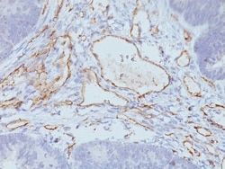
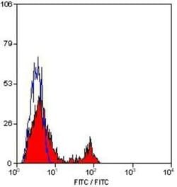
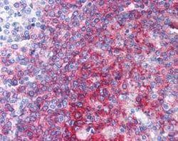
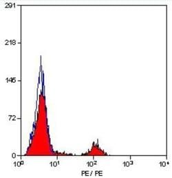
Flow Cytometry, Immunohistochemistry, Immunohistochemistry (Paraffin), Immunofluorescence
Conjugate
Unconjugated
Host Species
Mouse
Research Discipline
Apoptosis, Cell Cycle and Replication, Embryonic Stem Cell Markers, Immunology, Stem Cell Markers
Formulation
No buffer with 0.05% Sodium Azide
Gene Alias
CD30, CD30 antigen, CD30KI-1, CD30L receptor, cytokine receptor CD30, D1S166EKi-1, Ki-1 antigen, Lymphocyte activation antigen CD30, tumor necrosis factor receptor superfamily member 8, tumor necrosis factor receptor superfamily, member 8
Gene Symbols
TNFRSF8
Isotype
IgG1 κ
Purification Method
Tissue culture supernatant
Test Specificity
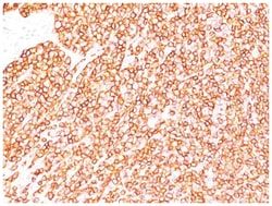
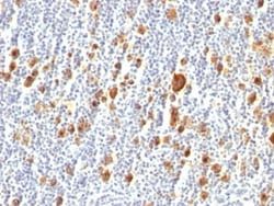
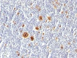
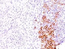
Recognizes a single chain glycoprotein of 105/120kDa, identified as CD30/Ki-1. Its epitope is located between aa112-412. CD30 is synthesized as a 90kDa precursor, which is processed in the Golgi complex into a membrane-bound phosphorylated mature 105/120kDa glycoprotein. In Hodgkin s disease, CD30/Ki-1 antigen is expressed by mononuclear-Hodgkin and multinucleated Reed-Sternberg cells. It is also expressed by the tumor cells of a majority of anaplastic large cell lymphomas as well as by a varying proportion of activated T and B cells. This MAb distinguishes large cell lymphomas derived from activated lymphoid cells from histiocytic malignancies and lymphomas derived from resting and precursor lymphoid cells or from anaplastic carcinomas. About one third of the Ki-1 positive lymphomas lack the leukocyte common antigen (CD45).
Clone
Ber-H2
Dilution
Flow Cytometry 5 - 10 ul/million cells in 0.1ml, Immunohistochemistry, Immunohistochemistry-Paraffin 1:50 - 1:100, Immunofluorescence 1:50 - 1:100
Classification
Monoclonal
Form
Supernatant
Regulatory Status
RUO
Target Species
Human
Gene Accession No.
P28908, P28908
Gene ID (Entrez)
943
Immunogen
Co cell line established from a patient with Hodgkin s disease of T-cell lineage
Primary or Secondary
Primary
Content And Storage
Store at 4C.
Related Products

Description
- Ensure accurate, reproducible results in Flow Cytometry, Immunohistochemistry (Paraffin), Immunofluorescence CD30/TNFRSF8 Monoclonal specifically detects CD30/TNFRSF8 in Human samples
- It is validated for Immunohistochemistry, Immunohistochemistry-Paraffin.
Compare Similar Items
Show Difference
Antigen: CD30/TNFRSF8
Concentration: 0.2mg/mL
Applications: Flow Cytometry, Immunohistochemistry, Immunohistochemistry (Paraffin), Immunofluorescence
Conjugate: Unconjugated
Host Species: Mouse
Research Discipline: Apoptosis, Cell Cycle and Replication, Embryonic Stem Cell Markers, Immunology, Stem Cell Markers
Formulation: No buffer with 0.05% Sodium Azide
Gene Alias: CD30, CD30 antigen, CD30KI-1, CD30L receptor, cytokine receptor CD30, D1S166EKi-1, Ki-1 antigen, Lymphocyte activation antigen CD30, tumor necrosis factor receptor superfamily member 8, tumor necrosis factor receptor superfamily, member 8
Gene Symbols: TNFRSF8
Isotype: IgG1 κ
Purification Method: Tissue culture supernatant
Test Specificity: Recognizes a single chain glycoprotein of 105/120kDa, identified as CD30/Ki-1. Its epitope is located between aa112-412. CD30 is synthesized as a 90kDa precursor, which is processed in the Golgi complex into a membrane-bound phosphorylated mature 105/120kDa glycoprotein. In Hodgkin s disease, CD30/Ki-1 antigen is expressed by mononuclear-Hodgkin and multinucleated Reed-Sternberg cells. It is also expressed by the tumor cells of a majority of anaplastic large cell lymphomas as well as by a varying proportion of activated T and B cells. This MAb distinguishes large cell lymphomas derived from activated lymphoid cells from histiocytic malignancies and lymphomas derived from resting and precursor lymphoid cells or from anaplastic carcinomas. About one third of the Ki-1 positive lymphomas lack the leukocyte common antigen (CD45).
Clone: Ber-H2
Dilution: Flow Cytometry 5 - 10 ul/million cells in 0.1ml, Immunohistochemistry, Immunohistochemistry-Paraffin 1:50 - 1:100, Immunofluorescence 1:50 - 1:100
Classification: Monoclonal
Form: Supernatant
Regulatory Status: RUO
Target Species: Human
Gene Accession No.: P28908, P28908
Gene ID (Entrez): 943
Immunogen: Co cell line established from a patient with Hodgkin s disease of T-cell lineage
Primary or Secondary: Primary
Content And Storage: Store at 4C.
Antigen: CD30/TNFRSF8
Concentration: 0.2mg/mL
Applications: Flow Cytometry, Immunohistochemistry (Paraffin), SDS-Page, Immunofluorescence
Conjugate: Unconjugated
Host Species: Mouse
Research Discipline: Apoptosis, Cell Cycle and Replication, Embryonic Stem Cell Markers, Immunology, Stem Cell Markers
Formulation: 10mM PBS and 0.05% BSA with 0.05% Sodium Azide
Gene Alias: CD30, CD30 antigen, CD30KI-1, CD30L receptor, cytokine receptor CD30, D1S166EKi-1, Ki-1 antigen, Lymphocyte activation antigen CD30, tumor necrosis factor receptor superfamily member 8, tumor necrosis factor receptor superfamily, member 8
Gene Symbols: TNFRSF8
Isotype: IgG1 κ
Purification Method: Protein A or G purified
Test Specificity: Recognizes a single chain glycoprotein of 105/120kDa, identified as CD30/Ki-1. CD30 is synthesized as a 90kDa precursor, which is processed in the Golgi complex into a membrane-bound phosphorylated mature 105/120kDa glycoprotein. In Hodgkins disease, CD30/Ki-1 antigen is expressed by mononuclear-Hodgkin and multinucleated Reed-Sternberg cells. It is also expressed by the tumor cells of a majority of anaplastic large cell lymphomas as well as by a varying proportion of activated T and B cells. This MAb distinguishes large cell lymphomas derived from activated lymphoid cells from histiocytic malignancies and lymphomas derived from resting and precursor lymphoid cells or from anaplastic carcinomas. About one third of the Ki-1 positive lymphomas lack the leukocyte common antigen (CD45).
Clone: Ki-1/779
Dilution: Flow Cytometry 0.5 - 1 ug/million cells in 0.1 ml, Immunohistochemistry-Paraffin 0.5 - 1.0 ug/ml, SDS-Page, Immunofluorescence 1 - 2 ug/ml
Classification: Monoclonal
Form: Purified
Regulatory Status: RUO
Target Species: Human
Gene Accession No.: P28908
Gene ID (Entrez): 943
Immunogen: Recombinant human TNFRSF8 protein
Primary or Secondary: Primary
Content And Storage: Store at 4C.
Antigen: CD30/TNFRSF8
Concentration: 0.2mg/mL
Applications: Flow Cytometry, Immunohistochemistry (Paraffin), Immunofluorescence
Conjugate: Unconjugated
Host Species: Mouse
Research Discipline: Apoptosis, Cell Cycle and Replication, Embryonic Stem Cell Markers, Immunology, Stem Cell Markers
Formulation: 10mM PBS and 0.05% BSA with 0.05% Sodium Azide
Gene Alias: CD30, CD30 antigen, CD30KI-1, CD30L receptor, cytokine receptor CD30, D1S166EKi-1, Ki-1 antigen, Lymphocyte activation antigen CD30, tumor necrosis factor receptor superfamily member 8, tumor necrosis factor receptor superfamily, member 8
Gene Symbols: TNFRSF8
Isotype: IgG1 κ
Purification Method: Protein A or G purified
Test Specificity: Recognizes a single chain glycoprotein of 105/120kDa, identified as CD30/Ki-1. CD30 is synthesized as a 90kDa precursor, which is processed in the Golgi complex into a membrane-bound phosphorylated mature 105/120kDa glycoprotein. In Hodgkin s disease, CD30/Ki-1 antigen is expressed by mononuclear-Hodgkin and multinucleated Reed-Sternberg cells. It is also expressed by the tumor cells of a majority of anaplastic large cell lymphomas as well as by a varying proportion of activated T and B cells. This MAb distinguishes large cell lymphomas derived from activated lymphoid cells from histiocytic malignancies and lymphomas derived from resting and precursor lymphoid cells or from anaplastic carcinomas. About one third of the Ki-1 positive lymphomas lack the leukocyte common antigen (CD45).
Clone: SPM609
Dilution: Flow Cytometry 0.5 - 1 ug/million cells in 0.1 ml, Immunohistochemistry-Paraffin 0.5 - 1.0 ug/ml, Immunofluorescence 1 - 2 ug/ml
Classification: Monoclonal
Form: Purified
Regulatory Status: RUO
Target Species: Human
Gene Accession No.: P28908
Gene ID (Entrez): 943
Immunogen: Recombinant human TNFRSF8 protein
Primary or Secondary: Primary
Content And Storage: Store at 4C.