CD36 Antibody (GPIIIb/1654), Novus Biologicals™
Mouse Monoclonal Antibody
Manufacturer: Fischer Scientific
The price for this product is unavailable. Please request a quote
Antigen
CD36/SR-B3
Concentration
0.2 mg/mL
Applications
Flow Cytometry, Immunohistochemistry (Paraffin), Immunofluorescence
Conjugate
Unconjugated
Host Species
Mouse
Research Discipline
Cancer, Cellular Markers, Endothelial Cell Markers, Hematopoietic Stem Cell Markers, Immunology, Lipid and Metabolism, Stem Cell Markers
Formulation
10mM PBS and 0.05% BSA with 0.05% Sodium Azide
Gene ID (Entrez)
948
Immunogen
Recombinant human GPIIIb protein
Primary or Secondary
Primary
Content And Storage
Store at 4C.
Molecular Weight of Antigen
85 kDa
Clone
GPIIIb/654
Dilution
Flow Cytometry 0.5 - 1 ug/million cells in 0.1 ml, Immunohistochemistry-Paraffin 0.5 - 1.0 ug/ml, Immunofluorescence 0.5 - 1.0 ug/ml
Classification
Monoclonal
Form
Purified
Regulatory Status
RUO
Target Species
Human
Gene Alias
CD36 antigen, CD36 molecule (thrombospondin receptor), FATCHDS7, Fatty acid translocase, Glycoprotein IIIb, GP3Bthrombospondin receptor), GPIIIB, Leukocyte differentiation antigen CD36, PAS IV, PAS-4, Platelet collagen receptor, platelet glycoprotein 4, Platelet glycoprotein IV, scavenger receptor class B, member 3, Thrombospondin receptor
Gene Symbols
CD36
Isotype
IgG2a κ
Purification Method
Protein A or G purified
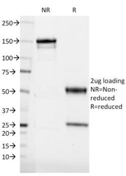
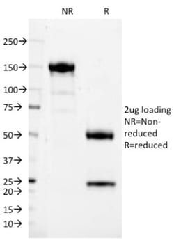
Test Specificity
Recognizes a protein of 80kDa-90kDa, identified as CD36. It is expressed on platelets, monocytes and macrophages, microvascular endothelial cells, erythrocyte precursors, mammary epithelial cells, and some macrophage derived dendritic cells. CD36 acts as a receptor for thrombospondin (TSP), collagen types I, IV and V, P. falciparum malaria-infected erythrocytes, and sickle erythrocytes. It also functions as a scavenger receptor, mediating macrophage uptake of oxidized low-density lipoprotein (LDL) and recognition of apoptotic polymorphonuclear leukocytes (PMN). CD36 plays a role in platelet aggregation, macrophage foam cell development, inflammation, and the tissue ischemia observed in sickle cell disease and cerebral malaria. Note that 1-4% of Japanese and East Asia population lack CD36.
Related Products


Description
- Ensure accurate, reproducible results in Flow Cytometry, Immunohistochemistry (Paraffin), Immunofluorescence CD36 Monoclonal specifically detects CD36 in Human samples
- It is validated for Flow Cytometry, Immunocytochemistry/Immunofluorescence, Immunofluorescence.
Compare Similar Items
Show Difference
Antigen: CD36/SR-B3
Concentration: 0.2 mg/mL
Applications: Flow Cytometry, Immunohistochemistry (Paraffin), Immunofluorescence
Conjugate: Unconjugated
Host Species: Mouse
Research Discipline: Cancer, Cellular Markers, Endothelial Cell Markers, Hematopoietic Stem Cell Markers, Immunology, Lipid and Metabolism, Stem Cell Markers
Formulation: 10mM PBS and 0.05% BSA with 0.05% Sodium Azide
Gene ID (Entrez): 948
Immunogen: Recombinant human GPIIIb protein
Primary or Secondary: Primary
Content And Storage: Store at 4C.
Molecular Weight of Antigen: 85 kDa
Clone: GPIIIb/654
Dilution: Flow Cytometry 0.5 - 1 ug/million cells in 0.1 ml, Immunohistochemistry-Paraffin 0.5 - 1.0 ug/ml, Immunofluorescence 0.5 - 1.0 ug/ml
Classification: Monoclonal
Form: Purified
Regulatory Status: RUO
Target Species: Human
Gene Alias: CD36 antigen, CD36 molecule (thrombospondin receptor), FATCHDS7, Fatty acid translocase, Glycoprotein IIIb, GP3Bthrombospondin receptor), GPIIIB, Leukocyte differentiation antigen CD36, PAS IV, PAS-4, Platelet collagen receptor, platelet glycoprotein 4, Platelet glycoprotein IV, scavenger receptor class B, member 3, Thrombospondin receptor
Gene Symbols: CD36
Isotype: IgG2a κ
Purification Method: Protein A or G purified
Test Specificity: Recognizes a protein of 80kDa-90kDa, identified as CD36. It is expressed on platelets, monocytes and macrophages, microvascular endothelial cells, erythrocyte precursors, mammary epithelial cells, and some macrophage derived dendritic cells. CD36 acts as a receptor for thrombospondin (TSP), collagen types I, IV and V, P. falciparum malaria-infected erythrocytes, and sickle erythrocytes. It also functions as a scavenger receptor, mediating macrophage uptake of oxidized low-density lipoprotein (LDL) and recognition of apoptotic polymorphonuclear leukocytes (PMN). CD36 plays a role in platelet aggregation, macrophage foam cell development, inflammation, and the tissue ischemia observed in sickle cell disease and cerebral malaria. Note that 1-4% of Japanese and East Asia population lack CD36.
Antigen: CD37
Concentration: 0.2 mg/mL
Applications: Flow Cytometry, SDS-Page, Immunofluorescence
Conjugate: Unconjugated
Host Species: Mouse
Research Discipline: Cellular Markers, Immunology
Formulation: 10mM PBS and 0.05% BSA with 0.05% Sodium Azide
Gene ID (Entrez): 951
Immunogen: Spleen cells of a patient with hairy cell leukemia
Primary or Secondary: Primary
Content And Storage: Store at 4C.
Molecular Weight of Antigen: __
Clone: IPO-24
Dilution: Flow Cytometry 0.5 - 1 ug/million cells in 0.1 ml, SDS-Page, Immunofluorescence 0.5 - 1.0 ug/ml
Classification: Monoclonal
Form: Purified
Regulatory Status: RUO
Target Species: Human
Gene Alias: CD37 antigentetraspanin-26, CD37 molecule, cell differentiation antigen 37, GP52-40, leukocyte antigen CD37, MGC120234, Tetraspanin-26, Tspan-26, TSPAN26leukocyte surface antigen CD37
Gene Symbols: CD37
Isotype: IgG2b κ
Purification Method: Protein A or G purified
Test Specificity: Recognizes a protein of 33-55kDa, identified as CD37 (Workshop V; Code CD37.7). CD37 is strongly expressed on normal and neoplastic mature (sIg+) B-lymphocytes. In B-cell ontogeny, CD37 appears after the pre-B-cell stage, is maintained during peripheral B-cell development and is lost upon terminal differentiation into plasma cells.1 CD37 is also present, at low densities, on resting and activated T cells, neutrophils, monocytes, and some myelomonocytic leukemia cells. It is absent from platelets, erythrocytes. CD37 is a member of a family of tetraspan transmembrane proteins, including CD9, CD53, CD63, CD81, and CD82. It associates other tetraspan transmembrane proteins and MHC class II molecules to form a large complex at the surface of B cells and play a role in signal transduction. CD37 is a valuable and stable marker for peripheral mature B-cells and corresponding malignancies like B-cell chronic lymphocytic leukemia (B-CLL), hairy cell leukemia (HCL), and all types of B-cell non-Ho