CD43/Sialophorin Antibody (SPN/839), Novus Biologicals™
Mouse Monoclonal Antibody
Manufacturer: Fischer Scientific
The price for this product is unavailable. Please request a quote
Antigen
CD43/Sialophorin
Concentration
0.2mg/mL
Applications
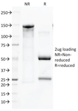
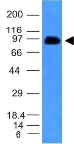
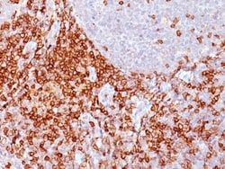
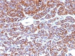
Western Blot, Flow Cytometry, Immunohistochemistry (Paraffin), SDS-Page, Immunofluorescence
Conjugate
Unconjugated
Host Species
Mouse
Research Discipline
Immunology
Formulation
10mM PBS and 0.05% BSA with 0.05% Sodium Azide
Gene Alias
CD43 antigen, CD43), Galactoglycoprotein, GALGP, Leukocyte sialoglycoprotein, Sialophorin, sialophorin (gpL115, leukosialin, CD43)
Gene Symbols
SPN
Isotype
IgG1 κ
Purification Method
Protein A or G purified
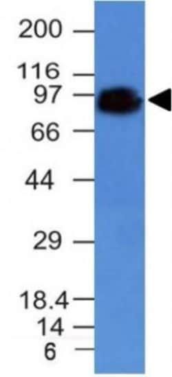
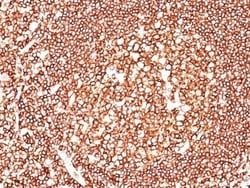
Test Specificity
It recognizes a cell surface glycoprotein of 95/115/135kDa (depending upon the extent of glycosylation), identified as CD43. 70-90% of T-cell lymphomas and from 22-37% of B-cell lymphomas express CD43. No reactivity has been observed with reactive B-cells. So a B-lineage population that co-expresses CD43 is highly likely to be a malignant lymphoma, especially a low-grade lymphoma, rather than a reactive B-cell population. When CD43 antibody is used in combination with anti-CD20, effective immunophenotyping of the lymphomas in formalin-fixed tissues can be obtained. Co-staining of a lymphoid infiltrate with anti-CD20 and anti-CD43 argues against a reactive process and favors a diagnosis of lymphoma.
Clone
SPN/839
Dilution
Western Blot 0.5 - 1.0 ug/ml, Flow Cytometry 0.5 - 1 ug/million cells in 0.1 ml, Immunohistochemistry-Paraffin 0.5 - 1.0 ug/ml, SDS-Page, Immunofluorescence 1 - 2 ug/ml
Classification
Monoclonal
Form
Purified
Regulatory Status
RUO
Target Species
Human
Gene Accession No.
P16150
Gene ID (Entrez)
6693
Immunogen
Recombinant full-length human SPN protein
Primary or Secondary
Primary
Content And Storage
Store at 4C.
Related Products

Description
- Ensure accurate, reproducible results in Western Blot, Flow Cytometry, Immunohistochemistry (Paraffin), Immunofluorescence CD43/Sialophorin Monoclonal specifically detects CD43/Sialophorin in Human samples
- It is validated for Western Blot, Flow Cytometry, Immunohistochemistry, Immunocytochemistry/Immunofluorescence, Immunohistochemistry-Paraffin.
Compare Similar Items
Show Difference
Antigen: CD43/Sialophorin
Concentration: 0.2mg/mL
Applications: Western Blot, Flow Cytometry, Immunohistochemistry (Paraffin), SDS-Page, Immunofluorescence
Conjugate: Unconjugated
Host Species: Mouse
Research Discipline: Immunology
Formulation: 10mM PBS and 0.05% BSA with 0.05% Sodium Azide
Gene Alias: CD43 antigen, CD43), Galactoglycoprotein, GALGP, Leukocyte sialoglycoprotein, Sialophorin, sialophorin (gpL115, leukosialin, CD43)
Gene Symbols: SPN
Isotype: IgG1 κ
Purification Method: Protein A or G purified
Test Specificity: It recognizes a cell surface glycoprotein of 95/115/135kDa (depending upon the extent of glycosylation), identified as CD43. 70-90% of T-cell lymphomas and from 22-37% of B-cell lymphomas express CD43. No reactivity has been observed with reactive B-cells. So a B-lineage population that co-expresses CD43 is highly likely to be a malignant lymphoma, especially a low-grade lymphoma, rather than a reactive B-cell population. When CD43 antibody is used in combination with anti-CD20, effective immunophenotyping of the lymphomas in formalin-fixed tissues can be obtained. Co-staining of a lymphoid infiltrate with anti-CD20 and anti-CD43 argues against a reactive process and favors a diagnosis of lymphoma.
Clone: SPN/839
Dilution: Western Blot 0.5 - 1.0 ug/ml, Flow Cytometry 0.5 - 1 ug/million cells in 0.1 ml, Immunohistochemistry-Paraffin 0.5 - 1.0 ug/ml, SDS-Page, Immunofluorescence 1 - 2 ug/ml
Classification: Monoclonal
Form: Purified
Regulatory Status: RUO
Target Species: Human
Gene Accession No.: P16150
Gene ID (Entrez): 6693
Immunogen: Recombinant full-length human SPN protein
Primary or Secondary: Primary
Content And Storage: Store at 4C.
Antigen: CD44
Concentration: 0.2mg/mL
Applications: Western Blot, Flow Cytometry, Immunohistochemistry (Paraffin), Immunofluorescence
Conjugate: Unconjugated
Host Species: Mouse
Research Discipline: Cancer, Cell Biology, Extracellular Matrix, Hematopoietic Stem Cell Markers, Immunology, Mesenchymal Stem Cell Markers, Signal Transduction, Stem Cell Markers
Formulation: 10mM PBS and 0.05% BSA with 0.05% Sodium Azide
Gene Alias: CD44 antigen, CD44 molecule (Indian blood group), CD44R, CDw44, cell surface glycoprotein CD44, chondroitin sulfate proteoglycan 8, CSPG8, ECMR-III, Epican, Extracellular matrix receptor III, GP90 lymphocyte homing/adhesion receptor, HCELL, hematopoietic cell E- and L-selectin ligand, Heparan sulfate proteoglycan, Hermes antigen, homing function and Indian blood group system, HUTCH-I, Hyaluronate receptor, IN, LHR, MC56, MDU2CD44 antigen (homing function and Indian blood group system), MDU3CDW44, MIC4MGC10468, Pgp1, PGP-1, PGP-I, Phagocytic glycoprotein 1, Phagocytic glycoprotein I
Gene Symbols: CD44
Isotype: IgG2a κ
Purification Method: Protein A or G purified
Test Specificity: Recognizes a cell surface glycoprotein of 80-95kDa (CD44) on lymphocytes, monocytes, and granulocytes (Leucocyte Typing Workshop V). Its epitope is resistant to digestion by trypsin and chymotrypsin. The CD44 family of glycoproteins exists in a number of variant isoforms, the most common being the standard 85-95kDa or hematopoietic variant (CD44s). Higher molecular weight isoforms are described in epithelial cells (CD44v), which are believed to function in intercellular adhesion and stromal binding. CD44 immunostaining is commonly used for the discrimination of urothelial transitional cell carcinoma in-situ from non-neoplastic changes in the urothelium.
Clone: HCAM/918
Dilution: Western Blot, Flow Cytometry 0.5 - 1 ug/million cells in 0.1 ml, Immunohistochemistry-Paraffin 0.25 - 0.5 ug/ml, Immunofluorescence 0.5 - 1.0 ug/ml
Classification: Monoclonal
Form: Purified
Regulatory Status: RUO
Target Species: Human, Primate
Gene Accession No.: __
Gene ID (Entrez): 960
Immunogen: Recombinant human HCAM protein
Primary or Secondary: Primary
Content And Storage: Store at 4C.
Antigen: CD45
Concentration: 0.2mg/mL
Applications: Flow Cytometry, Immunohistochemistry (Paraffin), SDS-Page, Immunofluorescence
Conjugate: Unconjugated
Host Species: Mouse
Research Discipline: Adaptive Immunity, Cell Biology, Cellular Markers, Cytokine Research, Glia Markers, Hematopoietic Stem Cell Markers, Immunology, Innate Immunity, MAP Kinase Signaling, Mast Cell Markers, Mesenchymal Stem Cell Markers, Microglia Markers, Modulation of DNA Pools, Myeloid Cell Markers, Neurodegeneration, Neuronal Cell Markers, Neuroscience, Signal Transduction, Stem Cell Markers
Formulation: 10mM PBS and 0.05% BSA with 0.05% Sodium Azide
Gene Alias: B220, CD45 antigen, CD45R, EC 3.1.3.48, L-CA, LY5, protein tyrosine phosphatase, receptor type, C, receptor-type tyrosine-protein phosphatase C, T200 glycoprotein, T200 leukocyte common antigen, T200receptor type, c polypeptide
Gene Symbols: PTPRC
Isotype: IgG2b κ
Purification Method: Protein A or G purified
Test Specificity: CD45R, also designated CD45 and PTPRC, has been identified as a transmembrane glycoprotein, broadly expressed among hematopoietic cells. Multiple isoforms of CD45R are distributed throughout the immune system according to cell type. These isoforms arise because of alternative splicing of exons 4, 5, and 6. The corresponding protein domains are characterized by the binding of monoclonal antibodies specific for CD45RA (exon 4), CD45RB (exon 5), CD45RC (exon 6) and CD45RO (exons 4 to 6 spliced out). The variation in these isoforms is localized to the extracellular domain of CD45R, while the intracellular domain is conserved. CD45R functions as a phosphor-tyrosine phosphatase. This MAb reacts with all isoforms of CD45R expressed by all hematopoietic cells, except erythrocytes, having a higher level of expression on lymphocytes than on granulocytes (Workshop IV). Antibody to CD45 is useful in differential diagnosis of lymphoid tumors from non-hematopoietic undifferentiated neoplasms.
Clone: 135-4C5
Dilution: Flow Cytometry 0.5 - 1 ug/million cells in 0.1 ml, Immunohistochemistry-Paraffin 0.5 - 1.0 ug/ml, SDS-Page, Immunofluorescence 0.5 - 1.0 ug/ml
Classification: Monoclonal
Form: Purified
Regulatory Status: RUO
Target Species: Human
Gene Accession No.: __
Gene ID (Entrez): 5788
Immunogen: Stimulated human leukocytes
Primary or Secondary: Primary
Content And Storage: Store at 4C.