Lewis A Blood Group Antigen Antibody (SPM279), Novus Biologicals™
Mouse Monoclonal Antibody
Manufacturer: Fischer Scientific
The price for this product is unavailable. Please request a quote
Antigen
Lewis A Blood Group Antigen
Concentration
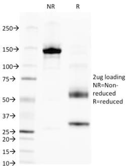
0.2mg/mL
Applications
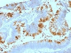
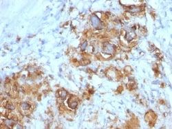
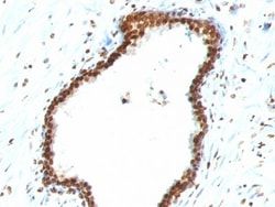
Flow Cytometry, Immunohistochemistry (Paraffin), Immunofluorescence
Conjugate
Unconjugated
Host Species
Mouse
Target Species
Human, Mouse
Gene ID (Entrez)
28
Immunogen
Mucins isolated from ovarian cyst fluid
Primary or Secondary
Primary
Content And Storage
Store at 4C.
Clone
SPM279
Dilution
Flow Cytometry 0.5 - 1 ug/million cells in 0.1 ml, Immunohistochemistry-Paraffin 0.5 - 1.0 ug/ml, Immunofluorescence 0.5 - 1.0 ug/ml
Classification
Monoclonal
Form
Purified
Regulatory Status
RUO
Formulation
10mM PBS and 0.05% BSA with 0.05% Sodium Azide
Gene Symbols
ABO
Isotype
IgG1 κ
Purification Method
Protein A or G purified
Test Specificity
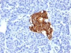
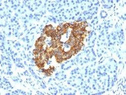
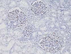
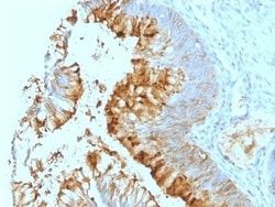
Recognizes a carbohydrate determinant of Gal 1-3(Fuc 1-4) GlcNAc which is blood group antigen Lewis A. It is present primarily on epithelial cells such as colon and kidneys. In the tumors and dedifferentiated tissues, decrease of Lewis A antigen was observed. Lewis A (type 1 chain) is expressed in colonic epithelial cells and may be useful for detection of gastrointestinal tumors, pancreatic cancer, and colorectal tumors. Blood group related antigens represent a group of carbohydrate determinants carried on both glycolipids and glycoproteins. They are usually mucin-type, and are detected on erythrocytes, certain epithelial cells, and in secretions of certain individuals. Sixteen genetically and biosynthetically distinct but inter-related specificities belong to this group of antigens, including A, B, H, Lewis A, Lewis B, Lewis X, Lewis Y, and precursor type 1 chain antigens.
Related Products
Description
- Ensure accurate, reproducible results in Flow Cytometry, Immunohistochemistry (Paraffin), Immunofluorescence Lewis A Blood Group Antigen Monoclonal specifically detects Lewis A Blood Group Antigen in Human, Mouse samples
- It is validated for Flow Cytometry, Immunohistochemistry, Immunocytochemistry/Immunofluorescence, Immunohistochemistry-Paraffin, Immunofluorescence.
Compare Similar Items
Show Difference
Antigen: Lewis A Blood Group Antigen
Concentration: 0.2mg/mL
Applications: Flow Cytometry, Immunohistochemistry (Paraffin), Immunofluorescence
Conjugate: Unconjugated
Host Species: Mouse
Target Species: Human, Mouse
Gene ID (Entrez): 28
Immunogen: Mucins isolated from ovarian cyst fluid
Primary or Secondary: Primary
Content And Storage: Store at 4C.
Clone: SPM279
Dilution: Flow Cytometry 0.5 - 1 ug/million cells in 0.1 ml, Immunohistochemistry-Paraffin 0.5 - 1.0 ug/ml, Immunofluorescence 0.5 - 1.0 ug/ml
Classification: Monoclonal
Form: Purified
Regulatory Status: RUO
Formulation: 10mM PBS and 0.05% BSA with 0.05% Sodium Azide
Gene Symbols: ABO
Isotype: IgG1 κ
Purification Method: Protein A or G purified
Test Specificity: Recognizes a carbohydrate determinant of Gal 1-3(Fuc 1-4) GlcNAc which is blood group antigen Lewis A. It is present primarily on epithelial cells such as colon and kidneys. In the tumors and dedifferentiated tissues, decrease of Lewis A antigen was observed. Lewis A (type 1 chain) is expressed in colonic epithelial cells and may be useful for detection of gastrointestinal tumors, pancreatic cancer, and colorectal tumors. Blood group related antigens represent a group of carbohydrate determinants carried on both glycolipids and glycoproteins. They are usually mucin-type, and are detected on erythrocytes, certain epithelial cells, and in secretions of certain individuals. Sixteen genetically and biosynthetically distinct but inter-related specificities belong to this group of antigens, including A, B, H, Lewis A, Lewis B, Lewis X, Lewis Y, and precursor type 1 chain antigens.
Antigen: Lewis A Blood Group Antigen
Concentration: 0.2mg/mL
Applications: Flow Cytometry, Immunohistochemistry (Paraffin), Immunofluorescence
Conjugate: Unconjugated
Host Species: Mouse
Target Species: Human, Mouse
Gene ID (Entrez): 28
Immunogen: Mucins isolated from ovarian cyst fluid
Primary or Secondary: Primary
Content And Storage: Store at 4C.
Clone: SPM522
Dilution: Flow Cytometry 0.5 - 1 ug/million cells in 0.1 ml, Immunohistochemistry-Paraffin 0.5 - 1.0 ug/ml, Immunofluorescence 0.5 - 1.0 ug/ml
Classification: Monoclonal
Form: Purified
Regulatory Status: RUO
Formulation: 10mM PBS and 0.05% BSA with 0.05% Sodium Azide
Gene Symbols: ABO
Isotype: IgG1 κ
Purification Method: Protein A or G purified
Test Specificity: Recognizes a carbohydrate determinant of Gal 1-3(Fuc 1-4) GlcNAc which is blood group antigen Lewis A. It is present primarily on epithelial cells such as colon and kidneys. In the tumors and dedifferentiated tissues, decrease of Lewis A antigen was observed. Lewis A (type 1 chain) is expressed in colonic epithelial cells and may be useful for detection of gastrointestinal tumors, pancreatic cancer, and colorectal tumors. Blood group related antigens represent a group of carbohydrate determinants carried on both glycolipids and glycoproteins. They are usually mucin-type, and are detected on erythrocytes, certain epithelial cells, and in secretions of certain individuals. Sixteen genetically and biosynthetically distinct but inter-related specificities belong to this group of antigens, including A, B, H, Lewis A, Lewis B, Lewis X, Lewis Y, and precursor type 1 chain antigens.

Antigen: Alkaline Phosphatase, Intestinal
Concentration: 0.2 mg/mL
Applications: Flow Cytometry, Immunohistochemistry (Frozen), Immunofluorescence
Conjugate: Unconjugated
Host Species: Mouse
Target Species: Human, Primate
Gene ID (Entrez): 248
Immunogen: Bovine intestinal alkaline phosphatase
Primary or Secondary: Primary
Content And Storage: Store at 4C.
Clone: SPM372
Dilution: Flow Cytometry 0.5 - 1 ug/million cells in 0.1 ml, Immunohistochemistry-Frozen 0.5 - 1.0 ug/ml, Immunofluorescence 0.5 - 1.0 ug/ml
Classification: Monoclonal
Form: Purified
Regulatory Status: RUO
Formulation: 10mM PBS and 0.05% BSA with 0.05% Sodium Azide
Gene Symbols: ALPI
Isotype: IgG1 κ
Purification Method: Protein A or G purified
Test Specificity: There are at least four distinct but related alkaline phosphatases: intestinal, placental, placental-like, and liver/bone/kidney (tissue non-specific). The first three are located together on chromosome 2, while the tissue non-specific form is located on chromosome 1. The product of this gene is a membrane bound glycosylated enzyme that is not expressed in any particular tissue and is, therefore, referred to as the tissue-nonspecific form of the enzyme. The exact physiological function of the alkaline phosphatases is not known. A proposed function of this form of the enzyme is matrix mineralization; however, mice that lack a functional form of this enzyme show normal skeletal development. This enzyme has been linked directly to hypo-phosphatasia, a disorder that is characterized by hypercalcemia and includes skeletal defects. The character of this disorder can vary, however, depending on the specific mutation since this determines age of onset and severity of symptoms. Alternatively spliced transcript variants, which encode the same protein, have been identified for this gene.