Arginase 1/ARG1/liver Arginase Antibody (ARG1/1125), Novus Biologicals™
Mouse Monoclonal Antibody
Manufacturer: Fischer Scientific
The price for this product is unavailable. Please request a quote
Antigen
Arginase 1/ARG1/liver Arginase
Concentration
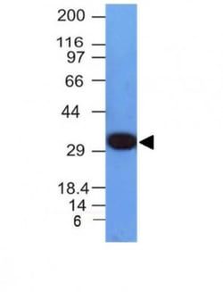
0.2mg/mL
Applications
Western Blot, Flow Cytometry, Immunohistochemistry (Paraffin), Immunofluorescence
Conjugate
Unconjugated
Host Species
Mouse
Research Discipline
Cancer, Cellular Markers, Chromatin Research, Lipid and Metabolism
Formulation
10mM PBS and 0.05% BSA with 0.05% Sodium Azide
Gene Alias
arginase 1, arginase, liver, arginase-1, EC 3.5.3.1, Liver-type arginase, Type I arginase
Gene Symbols
ARG1
Isotype
IgG3 κ
Purification Method
Protein A or G purified
Test Specificity
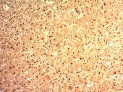
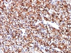
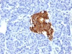
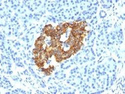
Recognizes a protein of 35-38kDa, which is identified as Arginase 1 (ARG1). Arginase is a manganese metallo-enzyme that catalyzes the hydrolysis of arginine to generate ornithine and urea. Arginase I and II are isoenzymes which differ in subcellular localization, regulation, and possibly function. Arginase I is a cytosolic enzyme, which is expressed mainly in the liver as part of the urea cycle, whereas arginase II is a mitochondrial protein found in a variety of tissues. Antibody to ARG-1 labels hepatocytes in normal tissues and granulocytes in peripheral blood. ARG-1 is a sensitive and specific marker for identification of hepatocellular carcinoma.
Clone
ARG1/1125
Dilution
Western Blot 0.5 - 1.0 ug/ml, Flow Cytometry 0.5 - 1 ug/million cells in 0.1 ml, Immunohistochemistry-Paraffin 2 - 4 ug/ml, Immunofluorescence 1 - 2 ug/ml
Classification
Monoclonal
Form
Purified
Regulatory Status
RUO
Target Species
Human
Gene Accession No.
P05089
Gene ID (Entrez)
383
Immunogen
Recombinant fragment (87 Amino acid residues around aa 1-150) of human ARG1 protein
Primary or Secondary
Primary
Content And Storage
Store at 4C.
Molecular Weight of Antigen
36.5 kDa
Related Products
Description
- Ensure accurate, reproducible results in Western Blot, Flow Cytometry, Immunohistochemistry (Paraffin), Immunofluorescence Arginase 1/ARG1/liver Arginase Monoclonal specifically detects Arginase 1/ARG1/liver Arginase in Human samples
- It is validated for Western Blot, Immunohistochemistry, Immunohistochemistry-Paraffin, Protein Array.
Compare Similar Items
Show Difference
Antigen: Arginase 1/ARG1/liver Arginase
Concentration: 0.2mg/mL
Applications: Western Blot, Flow Cytometry, Immunohistochemistry (Paraffin), Immunofluorescence
Conjugate: Unconjugated
Host Species: Mouse
Research Discipline: Cancer, Cellular Markers, Chromatin Research, Lipid and Metabolism
Formulation: 10mM PBS and 0.05% BSA with 0.05% Sodium Azide
Gene Alias: arginase 1, arginase, liver, arginase-1, EC 3.5.3.1, Liver-type arginase, Type I arginase
Gene Symbols: ARG1
Isotype: IgG3 κ
Purification Method: Protein A or G purified
Test Specificity: Recognizes a protein of 35-38kDa, which is identified as Arginase 1 (ARG1). Arginase is a manganese metallo-enzyme that catalyzes the hydrolysis of arginine to generate ornithine and urea. Arginase I and II are isoenzymes which differ in subcellular localization, regulation, and possibly function. Arginase I is a cytosolic enzyme, which is expressed mainly in the liver as part of the urea cycle, whereas arginase II is a mitochondrial protein found in a variety of tissues. Antibody to ARG-1 labels hepatocytes in normal tissues and granulocytes in peripheral blood. ARG-1 is a sensitive and specific marker for identification of hepatocellular carcinoma.
Clone: ARG1/1125
Dilution: Western Blot 0.5 - 1.0 ug/ml, Flow Cytometry 0.5 - 1 ug/million cells in 0.1 ml, Immunohistochemistry-Paraffin 2 - 4 ug/ml, Immunofluorescence 1 - 2 ug/ml
Classification: Monoclonal
Form: Purified
Regulatory Status: RUO
Target Species: Human
Gene Accession No.: P05089
Gene ID (Entrez): 383
Immunogen: Recombinant fragment (87 Amino acid residues around aa 1-150) of human ARG1 protein
Primary or Secondary: Primary
Content And Storage: Store at 4C.
Molecular Weight of Antigen: 36.5 kDa
Antigen: Gastric Mucin
Concentration: 0.2mg/mL
Applications: Flow Cytometry, Immunohistochemistry (Paraffin), Immunofluorescence
Conjugate: Unconjugated
Host Species: Mouse
Research Discipline: __
Formulation: 10mM PBS and 0.05% BSA with 0.05% Sodium Azide
Gene Alias: Gastric mucin-6, MUC-6, mucin 6, gastric, mucin 6, oligomeric mucus/gel-forming, mucin-6
Gene Symbols: MUC6
Isotype: IgG1 κ
Purification Method: Protein A or G purified
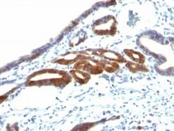
Test Specificity: The MUC6 gastric mucin is a secreted glycoprotein that plays an essential role in epithelial cyto-protection from acid, proteases, pathogenic microorganisms, and mechanical trauma in the gastrointestinal tract. Mucin 6 expression is highest in the stomach and gall bladder, with lower expression in the terminal ileum and right colon. In gastric cancer, Mucin 6 has an altered expression. In normal stomach, Mucin 6 is associated with Lewis type 2; Mucin 6 is also expressed in gastric metaplasia, duodenum and pancreas. Mucin 6 is a secretory mucin, located in the deeper mucosal folds of human gall bladder, and its expression is altered with increasing degrees of inflammation.
Clone: SPM598
Dilution: Flow Cytometry 0.5 - 1 ug/million cells in 0.1 ml, Immunohistochemistry-Paraffin 2 - 4 ug/ml, Immunofluorescence 1 - 2 ug/ml
Classification: Monoclonal
Form: Purified
Regulatory Status: RUO
Target Species: Human
Gene Accession No.: __
Gene ID (Entrez): 4588
Immunogen: A synthetic peptide of the Gastric Mucin 6 tandem repeat sequence.
Primary or Secondary: Primary
Content And Storage: Store at 4C.
Molecular Weight of Antigen: 252 kDa
Antigen: Gastric Mucin
Concentration: 0.2mg/mL
Applications: Flow Cytometry, Immunohistochemistry (Paraffin), Immunofluorescence
Conjugate: Unconjugated
Host Species: Mouse
Research Discipline: __
Formulation: 10mM PBS and 0.05% BSA with 0.05% Sodium Azide
Gene Alias: Gastric mucin-6, MUC-6, mucin 6, gastric, mucin 6, oligomeric mucus/gel-forming, mucin-6
Gene Symbols: MUC6
Isotype: IgG1 κ
Purification Method: Protein A or G purified
Test Specificity: The MUC6 gastric mucin is a secreted glycoprotein that plays an essential role in epithelial cyto-protection from acid, proteases, pathogenic microorganisms, and mechanical trauma in the gastrointestinal tract. Mucin 6 expression is highest in the stomach and gall bladder, with lower expression in the terminal ileum and right colon. In gastric cancer, Mucin 6 has an altered expression. In normal stomach, Mucin 6 is associated with Lewis type 2; Mucin 6 is also expressed in gastric metaplasia, duodenum and pancreas. Mucin 6 is a secretory mucin, located in the deeper mucosal folds of human gall bladder, and its expression is altered with increasing degrees of inflammation.
Clone: SPM598
Dilution: Flow Cytometry 0.5 - 1 ug/million cells in 0.1 ml, Immunohistochemistry-Paraffin 2 - 4 ug/ml, Immunofluorescence 1 - 2 ug/ml
Classification: Monoclonal
Form: Purified
Regulatory Status: RUO
Target Species: Human
Gene Accession No.: __
Gene ID (Entrez): 4588
Immunogen: A synthetic peptide of the Gastric Mucin 6 tandem repeat sequence.
Primary or Secondary: Primary
Content And Storage: Store at 4C.
Molecular Weight of Antigen: 252 kDa
