Napsin A Antibody (NAPSA/1238) - BSA Free, Novus Biologicals™
Mouse Monoclonal Antibody
Manufacturer: Fischer Scientific
The price for this product is unavailable. Please request a quote
Antigen
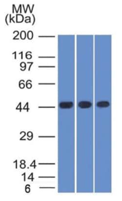
Napsin A
Concentration
1 mg/mL
Applications
Western Blot, Flow Cytometry, Immunocytochemistry, Immunofluorescence, Immunohistochemistry (Paraffin), CyTOF
Conjugate
Unconjugated
Host Species
Mouse
Research Discipline
Cancer, Cell Biology, Cellular Markers
Formulation
PBS with No Preservative
Gene ID (Entrez)
9476
Immunogen
Recombinant fragment of human Napsin-A protein
Primary or Secondary
Primary
Content And Storage
Store at -20 to -80C. Avoid freeze-thaw cycles.
Molecular Weight of Antigen
37 kDa
Clone
NAPSA/1238
Dilution
Western Blot 1 - 2 ug/ml, Flow Cytometry 0.5 - 1 ug/million cells, Immunocytochemistry/Immunofluorescence 1 - 2 ug/ml, Immunohistochemistry-Paraffin 1 - 2 ug/ml, CyTOF-ready
Classification
Monoclonal
Form
Purified
Regulatory Status
RUO
Target Species
Human
Gene Alias
Asp 4, ASP4, Aspartyl protease 4, EC 3.4.23, EC 3.4.23.-, EC 3.4.23.15, EC 3.4.23.3, EC 3.4.23.5, KAP, Kdap, NAP1napsin-A, NAPApronapsin A, NAPSA, napsin A aspartic peptidase, napsin-1, SNAPA, TA01/TA02
Gene Symbols
NAPSA
Isotype
IgG κ
Purification Method
Protein A purified
Test Specificity
Napsin is a pepsin-like aspartic proteinase connected with maturation of surfactant protein B.There are two closely related napsins, napsin A and napsin B. Napsin A is expressed as a single chain protein. Immunohistochemical studies revealed high expression levels of napsin A in human lung and kidney but low expression in spleen. Napsin A is expressed in type II pneumocytes and in adenocarcinomas of lung. The high specificity expression of napsin A in adenocarcinomas of lung is useful to distinguish primary lung adenocarcinomas from adenocarcinomas of other organs.
Description
- Napsin A Monoclonal specifically detects Napsin A in Human samples
- It is validated for Western Blot, Flow Cytometry, Immunohistochemistry, Immunocytochemistry/Immunofluorescence, Immunohistochemistry-Paraffin, Protein Array, CyTOF-ready.
Compare Similar Items
Show Difference
Antigen: Napsin A
Concentration: 1 mg/mL
Applications: Western Blot, Flow Cytometry, Immunocytochemistry, Immunofluorescence, Immunohistochemistry (Paraffin), CyTOF
Conjugate: Unconjugated
Host Species: Mouse
Research Discipline: Cancer, Cell Biology, Cellular Markers
Formulation: PBS with No Preservative
Gene ID (Entrez): 9476
Immunogen: Recombinant fragment of human Napsin-A protein
Primary or Secondary: Primary
Content And Storage: Store at -20 to -80C. Avoid freeze-thaw cycles.
Molecular Weight of Antigen: 37 kDa
Clone: NAPSA/1238
Dilution: Western Blot 1 - 2 ug/ml, Flow Cytometry 0.5 - 1 ug/million cells, Immunocytochemistry/Immunofluorescence 1 - 2 ug/ml, Immunohistochemistry-Paraffin 1 - 2 ug/ml, CyTOF-ready
Classification: Monoclonal
Form: Purified
Regulatory Status: RUO
Target Species: Human
Gene Alias: Asp 4, ASP4, Aspartyl protease 4, EC 3.4.23, EC 3.4.23.-, EC 3.4.23.15, EC 3.4.23.3, EC 3.4.23.5, KAP, Kdap, NAP1napsin-A, NAPApronapsin A, NAPSA, napsin A aspartic peptidase, napsin-1, SNAPA, TA01/TA02
Gene Symbols: NAPSA
Isotype: IgG κ
Purification Method: Protein A purified
Test Specificity: Napsin is a pepsin-like aspartic proteinase connected with maturation of surfactant protein B.There are two closely related napsins, napsin A and napsin B. Napsin A is expressed as a single chain protein. Immunohistochemical studies revealed high expression levels of napsin A in human lung and kidney but low expression in spleen. Napsin A is expressed in type II pneumocytes and in adenocarcinomas of lung. The high specificity expression of napsin A in adenocarcinomas of lung is useful to distinguish primary lung adenocarcinomas from adenocarcinomas of other organs.

Antigen: N-Cadherin
Concentration: 0.2 mg/mL
Applications: Western Blot, Flow Cytometry, Immunocytochemistry, Immunofluorescence, Immunohistochemistry (Paraffin)
Conjugate: Unconjugated
Host Species: Mouse
Research Discipline: Cancer, Cell Cycle and Replication, Cellular Markers, Extracellular Matrix, Phospho Specific, Plasma Membrane Markers, Signal Transduction
Formulation: 10mM PBS with 0.05% BSA with 0.05% Sodium Azide
Gene ID (Entrez): 1000
Immunogen: Recombinant human N-cadherin extracellular domain (exact sequence is proprietary)
Primary or Secondary: Primary
Content And Storage: Store at 4C.
Molecular Weight of Antigen: 135 kDa
Clone: 8C11
Dilution: Western Blot 0.5 - 1.0 ug/ml, Flow Cytometry 0.5 - 1 ug/million cells, Immunocytochemistry/Immunofluorescence 1 - 2 ug/ml, Immunohistochemistry-Paraffin 0.5 - 1.0 ug/ml
Classification: Monoclonal
Form: Purified
Regulatory Status: RUO
Target Species: Human, Mouse
Gene Alias: cadherin 2, N-cadherin (neuronal), cadherin 2, type 1, N-cadherin (neuronal), cadherin-2, CD325, CD325 antigen, CDHNcalcium-dependent adhesion protein, neuronal, CDw325, N-cadherin, NCADN-cadherin 1, Neural cadherin, neural-cadherin
Gene Symbols: CDH2
Isotype: IgG1 κ
Purification Method: Protein A or G purified
Test Specificity: __