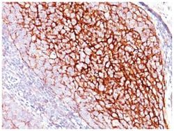
CD31/PECAM-1 Antibody (SPM532), Novus Biologicals™
Mouse Monoclonal Antibody
Manufacturer: Fischer Scientific
The price for this product is unavailable. Please request a quote
Antigen
CD31/PECAM-1
Concentration
0.2mg/mL
Applications
Flow Cytometry, Immunocytochemistry, Immunofluorescence, Immunohistochemistry (Paraffin)
Conjugate
Unconjugated
Host Species
Mouse
Research Discipline
Angiogenesis, Cancer, Cellular Markers, Cytoskeleton Markers, Embryonic Stem Cell Markers, Endothelial Cell Markers, Extracellular Matrix, Hematopoietic Stem Cell Markers, Immunology, Mesenchymal Stem Cell Markers, Myeloid Cell Markers, Signal Transduction, Stem Cell Markers
Formulation
10mM PBS and 0.05% BSA with 0.05% Sodium Azide
Gene Alias
adhesion molecule, CD31, CD31 antigen, CD31/EndoCAM, EndoCAM, FLJ34100, FLJ58394, GPIIA', PECA1, PECAM-1, PECAM-1, CD31/EndoCAM, platelet endothelial cell adhesion molecule, platelet/endothelial cell adhesion molecule
Gene Symbols
PECAM1
Isotype
IgG1 κ
Purification Method
Protein A or G purified
Test Specificity
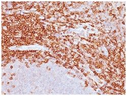
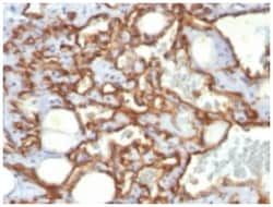
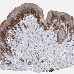
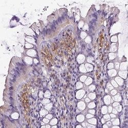
CD31 (PECAM-1) is a transmembrane glycoprotein member of the immunoglobulin supergene family of adhesion molecules. CD31 is expressed by stem cells of the hematopoietic system and is primarily used to identify and concentrate these cells for experimental studies as well as for bone marrow transplantation. Anti-CD31 has shown to be highly specific and sensitive for vascular endothelial cells. Staining of nonvascular tumors (excluding hematopoietic neoplasms) is rare. CD31 MAb reacts with normal, benign, and malignant endothelial cells which make up blood vessel lining. The level of CD31 expression can help to determine the degree of tumor angiogenesis, and a high level of CD31 expression may imply a rapidly growing tumor and potentially a predictor of tumor recurrence.
Clone
SPM532
Dilution
Flow Cytometry 0.5-1ug/million cells, Immunocytochemistry/Immunofluorescence 1-2ug/ml, Immunohistochemistry-Paraffin 0.5-1.0ug/ml
Classification
Monoclonal
Form
Purified
Regulatory Status
RUO
Target Species
Human
Gene Accession No.
P16284
Gene ID (Entrez)
5175
Immunogen
Human recombinant CD31 protein
Primary or Secondary
Primary
Content And Storage
Store at 4C.
Related Products
Description
- CD31/PECAM-1 Monoclonal specifically detects CD31/PECAM-1 in Human samples
- It is validated for Western Blot, Flow Cytometry, Immunohistochemistry, Immunocytochemistry/Immunofluorescence, Immunohistochemistry-Paraffin.
Compare Similar Items
Show Difference
Antigen: CD31/PECAM-1
Concentration: 0.2mg/mL
Applications: Flow Cytometry, Immunocytochemistry, Immunofluorescence, Immunohistochemistry (Paraffin)
Conjugate: Unconjugated
Host Species: Mouse
Research Discipline: Angiogenesis, Cancer, Cellular Markers, Cytoskeleton Markers, Embryonic Stem Cell Markers, Endothelial Cell Markers, Extracellular Matrix, Hematopoietic Stem Cell Markers, Immunology, Mesenchymal Stem Cell Markers, Myeloid Cell Markers, Signal Transduction, Stem Cell Markers
Formulation: 10mM PBS and 0.05% BSA with 0.05% Sodium Azide
Gene Alias: adhesion molecule, CD31, CD31 antigen, CD31/EndoCAM, EndoCAM, FLJ34100, FLJ58394, GPIIA', PECA1, PECAM-1, PECAM-1, CD31/EndoCAM, platelet endothelial cell adhesion molecule, platelet/endothelial cell adhesion molecule
Gene Symbols: PECAM1
Isotype: IgG1 κ
Purification Method: Protein A or G purified
Test Specificity: CD31 (PECAM-1) is a transmembrane glycoprotein member of the immunoglobulin supergene family of adhesion molecules. CD31 is expressed by stem cells of the hematopoietic system and is primarily used to identify and concentrate these cells for experimental studies as well as for bone marrow transplantation. Anti-CD31 has shown to be highly specific and sensitive for vascular endothelial cells. Staining of nonvascular tumors (excluding hematopoietic neoplasms) is rare. CD31 MAb reacts with normal, benign, and malignant endothelial cells which make up blood vessel lining. The level of CD31 expression can help to determine the degree of tumor angiogenesis, and a high level of CD31 expression may imply a rapidly growing tumor and potentially a predictor of tumor recurrence.
Clone: SPM532
Dilution: Flow Cytometry 0.5-1ug/million cells, Immunocytochemistry/Immunofluorescence 1-2ug/ml, Immunohistochemistry-Paraffin 0.5-1.0ug/ml
Classification: Monoclonal
Form: Purified
Regulatory Status: RUO
Target Species: Human
Gene Accession No.: P16284
Gene ID (Entrez): 5175
Immunogen: Human recombinant CD31 protein
Primary or Secondary: Primary
Content And Storage: Store at 4C.

Antigen: Siglec-3/CD33
Concentration: __
Applications: Flow Cytometry, Immunocytochemistry, Immunofluorescence, Immunoprecipitation, Immunohistochemistry (Frozen)
Conjugate: Unconjugated
Host Species: Mouse
Research Discipline: Immunology
Formulation: PBS with 0.05% BSA. with 0.05% Sodium Azide
Gene Alias: CD33 antigen, CD33 antigen (gp67), CD33 molecule, FLJ00391, myeloid cell surface antigen CD33, p67, sialic acid binding Ig-like lectin 3, Sialic acid-binding Ig-like lectin 3, SIGLEC-3, SIGLEC3gp67
Gene Symbols: CD33
Isotype: IgG1 κ
Purification Method: Protein A purified
Test Specificity: Recognizes a 67kDa glycoprotein, which is identified as CD33. It is a transmembrane protein of the sialic acid-binding immunoglobulin-like lectin (Siglec) family. It belongs to the immunoreceptor tyrosine-based inhibitory motif (ITIM)-containing molecules able of recruiting protein tyrosine phosphatases SHP-1 and SHP-2 to signal assemblies; these ITIMs are also used for ubiquitin-mediated removal of the receptor from the cell surface. CD33 is expressed on cells of myelomonocytic lineage, binds sialic acid residues in N- and O-glycans on cell surfaces, and is a therapeutic target for acute myeloid leukemia. CD33 is expressed on myeloid progenitors, monocytes, granulocytes, dendritic cells and mast cells. It is absent on platelets, lymphocytes, erythrocytes and hematopoietic stem cells.
Clone: C33/69
Dilution: Flow Cytometry 0.5-1ug/million cells, Immunocytochemistry/Immunofluorescence 0.5-1ug/ml, Immunoprecipitation 0.5-1ug/500ug protein lysate, Immunohistochemistry-Frozen 0.5-1ug/ml
Classification: Monoclonal
Form: Purified
Regulatory Status: RUO
Target Species: Human
Gene Accession No.: P20138
Gene ID (Entrez): 945
Immunogen: Recombinant human CD33 protein
Primary or Secondary: Primary
Content And Storage: Store at 4C.
Antigen: CD35
Concentration: __
Applications: Western Blot, Flow Cytometry, Immunocytochemistry, Immunofluorescence, Immunoprecipitation, Immunohistochemistry (Paraffin)
Conjugate: Unconjugated
Host Species: Mouse
Research Discipline: __
Formulation: PBS with 0.05% BSA. with 0.05% Sodium Azide
Gene Alias: complement component (3b/4b) receptor 1 (Knops blood group), KN
Gene Symbols: CR1
Isotype: IgG1 κ
Purification Method: Protein A purified
Test Specificity: Recognizes a protein of 210-220kDa, which is identified as the complement receptor 1 (CR1)/CD35. This MAb is specific for a site in CR1 that is distal from the C3b-binding site, so that it is unable to block CR1 activity. This MAb is highly specific to CR1 and shows no cross-reaction with CR2. The primary function of CR1 is to serve as the cellular receptor for C3b and C4b, the most important components of the complement system leading to clearance of foreign macromolecules. The Knops blood group system is a system of antigens located on this protein.Follicular dendritic cells (FDC) are restricted to the B-cell regions of secondary lymphoid follicles. They are CD21+/CD35+/CD1a-. This MAb labels follicular dendritic cells and follicular dendritic cell sarcoma.
Clone: SPM554
Dilution: Western Blot 0.5-1ug/ml, Flow Cytometry 0.5-1ug/million cells, Immunocytochemistry/Immunofluorescence 1-2ug/ml, Immunoprecipitation 0.5-1ug/500ug protein lysate, Immunohistochemistry-Paraffin 0.5-1.0ug/ml, Immunohistochemistry-Frozen 0.5-1.0ug/ml
Classification: Monoclonal
Form: Purified
Regulatory Status: RUO
Target Species: Human, Baboon, Primate
Gene Accession No.: P17927
Gene ID (Entrez): 1378
Immunogen: Intact human monocytes
Primary or Secondary: Primary
Content And Storage: Store at 4C.