MAGE 1 Mouse, Clone: SPM282, Novus Biologicals™
Mouse Monoclonal Antibody
Manufacturer: Fischer Scientific
The price for this product is unavailable. Please request a quote
Antigen
MAGE 1
Dilution
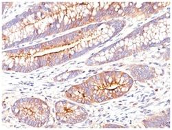
Flow Cytometry 0.5-1ug/million cells, Immunocytochemistry/Immunofluorescence 1-2ug/ml, Immunohistochemistry-Paraffin 0.5-1.0ug/ml
Classification
Monoclonal
Form
Purified
Regulatory Status
RUO
Target Species
Human, Rat, Canine
Gene Accession No.
P43355
Gene ID (Entrez)
4100
Immunogen
Human MAGE-A1 full length recombinant protein
Primary or Secondary
Primary
Content And Storage
Store at 4C.
Clone
SPM282
Applications
Flow Cytometry, Immunocytochemistry, Immunofluorescence, Immunohistochemistry (Paraffin)
Conjugate
Unconjugated
Host Species
Mouse
Research Discipline
Apoptosis, Cancer, Melanoma Cell Markers, Tumor Suppressors
Formulation
PBS with 0.05% BSA. with 0.05% Sodium Azide
Gene Alias
Antigen MZ2-E, Cancer/testis antigen 1.1, cancer/testis antigen family 1, member 1, CT1.1melanoma antigen family A 1, MAGE-1 antigen, MAGE1A, MAGE1melanoma antigen MAGE-1, melanoma antigen family A, 1 (directs expression of antigen MZ2-E), melanoma-associated antigen 1, melanoma-associated antigen MZ2-E, MGC9326
Gene Symbols
MAGEA1
Isotype
IgG1 κ
Purification Method
Protein A purified
Test Specificity
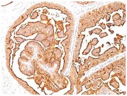
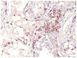
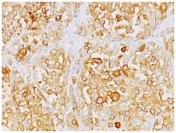
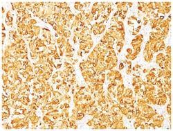
Recognizes a protein of 42-46kDa, identified as MAGE-1. This MAb does not cross-react with MAGE-2, -3, -4, -6 -9, -10, -or -12 protein. Human malignant neoplasms carry rejection antigens that are recognized by the patients' autologous, tumor directed and specific, cytolytic, CD8+ T lymphocyte clones (CTL). The MAGE family of genes codes an important group of antigens. It was identified that melanomas and primary glial brain tumors express common melanoma associated antigens (MAAs). Because MAGE-1 is expressed on a significant proportion of human neoplasms of various histological types (melanoma, brain tumors of glial origin, neuroblastoma, non-small cell lung cancer, breast, gastric, colorectal, ovarian, renal cell carcinomas) and not on normal tissues, the encoded antigen may serve as a marker of early detection and target for cancer immunotherapy.
Related Products


Description
- MAGE 1 Monoclonal specifically detects MAGE 1 in Human, Rat, Canine samples
- It is validated for Flow Cytometry, Immunohistochemistry, Immunocytochemistry/Immunofluorescence, Immunohistochemistry-Paraffin, Flow (Intracellular).
Compare Similar Items
Show Difference
Antigen: MAGE 1
Dilution: Flow Cytometry 0.5-1ug/million cells, Immunocytochemistry/Immunofluorescence 1-2ug/ml, Immunohistochemistry-Paraffin 0.5-1.0ug/ml
Classification: Monoclonal
Form: Purified
Regulatory Status: RUO
Target Species: Human, Rat, Canine
Gene Accession No.: P43355
Gene ID (Entrez): 4100
Immunogen: Human MAGE-A1 full length recombinant protein
Primary or Secondary: Primary
Content And Storage: Store at 4C.
Clone: SPM282
Applications: Flow Cytometry, Immunocytochemistry, Immunofluorescence, Immunohistochemistry (Paraffin)
Conjugate: Unconjugated
Host Species: Mouse
Research Discipline: Apoptosis, Cancer, Melanoma Cell Markers, Tumor Suppressors
Formulation: PBS with 0.05% BSA. with 0.05% Sodium Azide
Gene Alias: Antigen MZ2-E, Cancer/testis antigen 1.1, cancer/testis antigen family 1, member 1, CT1.1melanoma antigen family A 1, MAGE-1 antigen, MAGE1A, MAGE1melanoma antigen MAGE-1, melanoma antigen family A, 1 (directs expression of antigen MZ2-E), melanoma-associated antigen 1, melanoma-associated antigen MZ2-E, MGC9326
Gene Symbols: MAGEA1
Isotype: IgG1 κ
Purification Method: Protein A purified
Test Specificity: Recognizes a protein of 42-46kDa, identified as MAGE-1. This MAb does not cross-react with MAGE-2, -3, -4, -6 -9, -10, -or -12 protein. Human malignant neoplasms carry rejection antigens that are recognized by the patients' autologous, tumor directed and specific, cytolytic, CD8+ T lymphocyte clones (CTL). The MAGE family of genes codes an important group of antigens. It was identified that melanomas and primary glial brain tumors express common melanoma associated antigens (MAAs). Because MAGE-1 is expressed on a significant proportion of human neoplasms of various histological types (melanoma, brain tumors of glial origin, neuroblastoma, non-small cell lung cancer, breast, gastric, colorectal, ovarian, renal cell carcinomas) and not on normal tissues, the encoded antigen may serve as a marker of early detection and target for cancer immunotherapy.
Antigen: Moesin
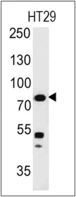
Dilution: Western Blot 0.5-1ug/ml, Flow Cytometry 0.5-1ug/million cells, Immunocytochemistry/Immunofluorescence 0.5-1ug/ml, Immunohistochemistry-Paraffin 0.5-1.0ug/ml
Classification: Monoclonal
Form: Purified
Regulatory Status: RUO
Target Species: Human, Rat
Gene Accession No.: P26038
Gene ID (Entrez): 4478
Immunogen: Recombinant human Moesin protein
Primary or Secondary: Primary
Content And Storage: Store at 4C.
Clone: SPM562
Applications: Western Blot, Flow Cytometry, Immunocytochemistry, Immunofluorescence, Immunohistochemistry (Paraffin)
Conjugate: Unconjugated
Host Species: Mouse
Research Discipline: Cytoskeleton Markers, Stem Cell Markers
Formulation: PBS with 0.05% BSA. with 0.05% Sodium Azide
Gene Alias: Membrane-organizing extension spike protein, moesin
Gene Symbols: MSN
Isotype: IgG1 κ
Purification Method: Protein A purified
Test Specificity: Recognizes 78kDa moesin protein. Moesin, a member of the talin-4.1 superfamily, is a linking protein of the sub-membranous actin cytoskeleton. It is expressed in variable amounts in cells of different phenotypes such as macrophages, lymphocytes, fibroblastic, endothelial, epithelial, and neuronal cell lines but not in blood cells. The ERM proteins, ezrin, radixin, and moesin are involved in a variety of cellular functions, such as cell adhesion, migration, and the organization of cell surface structures, and are highly homologous, both in protein sequence and in functional activity, with merlin/schwannomin, a neurofibromatosis-2-associated tumor-suppressor protein. Cell lines of epithelial and mesothelial origin contain both moesin and radixin whereas cells of endothelial and lymphoid origin express moesin.
Antigen: MUC-1
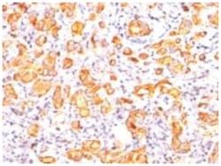
Dilution: Western Blot 0.5-1ug/ml, Flow Cytometry 0.5-1ug/million cells, Immunocytochemistry/Immunofluorescence 1-2ug/ml, Immunohistochemistry-Paraffin 0.5-1.0ug/ml
Classification: Monoclonal
Form: Purified
Regulatory Status: RUO
Target Species: Human
Gene Accession No.: P15941
Gene ID (Entrez): 4582
Immunogen: Human milk fat globule membranes
Primary or Secondary: Primary
Content And Storage: Store at 4C.
Clone: SPM132
Applications: Western Blot, Flow Cytometry, Immunocytochemistry, Immunofluorescence, Immunohistochemistry (Paraffin)
Conjugate: Unconjugated
Host Species: Mouse
Research Discipline: Cancer, Cellular Markers, Extracellular Matrix
Formulation: PBS with 0.05% BSA. with 0.05% Sodium Azide
Gene Alias: Breast carcinoma-associated antigen DF3, Carcinoma-associated mucin, CD227, CD227 antigen, DF3 antigen, EMA, episialin, H23 antigen, H23AG, KL-6, MAM6, MUC-1, MUC1/ZD, mucin 1, cell surface associated, mucin 1, transmembrane, mucin-1, Peanut-reactive urinary mucin, PEMMUC-1/SEC, PEMT, Polymorphic epithelial mucin, PUMMUC-1/X, tumor associated epithelial mucin, Tumor-associated epithelial membrane antigen, Tumor-associated mucin
Gene Symbols: MUC1
Isotype: IgG1 κ
Purification Method: Protein A purified
Test Specificity: In Western blotting, it recognizes proteins in MW range of 265-400kDa, identified as different glycoforms of EMA. This MAb reacts with the DTRP epitope in the tandem repeats. The alpha subunit has cell adhesive properties. It can act both as an adhesion and an anti-adhesion protein. EMA may provide a protective layer on epithelial cells against bacterial and enzyme attack. The beta subunit contains a C-terminal domain, which is involved in cell signaling, through phosphorylations and protein-protein interactions. In immunohistochemical assays, it superbly stains routine formalin/paraffin carcinoma tissues. Antibody to EMA is useful as a pan-epithelial marker for detecting early metastatic loci of carcinoma in bone marrow or liver.