alpha-Methylacyl-CoA Racemase/AMACR Antibody, Novus Biologicals™
Rabbit Polyclonal Antibody
Manufacturer: Fischer Scientific
The price for this product is unavailable. Please request a quote
Antigen
alpha-Methylacyl-CoA Racemase/AMACR
Applications
Western Blot, Immunohistochemistry (Paraffin)
Conjugate
Unconjugated
Host Species
Rabbit
Research Discipline
Lipid and Metabolism, Prostate Cancer, Signal Transduction
Formulation
PBS with 0.05% BSA. with 0.05% Sodium Azide
Gene Alias
2-methylacyl-CoA racemase, alpha-methylacyl-CoA racemase, CBAS4, EC 5.1.99.4, RACE, RM
Gene Symbols
AMACR
Isotype
IgG
Purification Method
Protein A purified
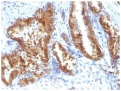
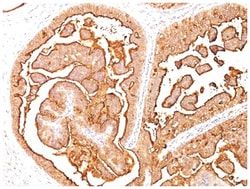
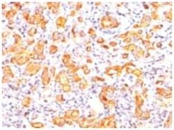
Test Specificity
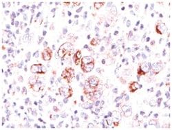
This antibody recognizes a protein of 54kDa, which is identified as AMACR, also known as p504S. It is an enzyme that is involved in bile acid biosynthesis and oxidation of branched-chain fatty acids. AMACR is essential in lipid metabolism. It is expressed in cells of premalignant high-grade prostatic intraepithelial neoplasia (HGPIN) and prostate adenocarcinoma. The majority of the carcinoma cells show a distinct granular cytoplasmic staining reaction. AMACR is present at low or undetectable levels in glandular epithelial cells of normal prostate and benign prostatic hyperplasia. A spotty granular cytoplasmic staining is seen in a few cells of the benign glands. AMACR is expressed in normal liver (hepatocytes), kidney (tubular epithelial cells) and gall bladder (epithelial cells). Expression has also been found in lung (bronchial epithelial cells) and colon (colonic surface epithelium). AMACR expression can also be found in hepatocellular carcinoma and kidney carcinoma. Past studies ha
Dilution
Western Blot 1:100-1:200, Immunohistochemistry-Paraffin 1:50-1:100
Classification
Polyclonal
Form
Purified
Regulatory Status
RUO
Target Species
Human
Gene Accession No.
Q9UHK6
Gene ID (Entrez)
23600
Immunogen
A synthetic peptide from human AMACR protein
Primary or Secondary
Primary
Content And Storage
Store at 4C.
Molecular Weight of Antigen
54 kDa
Related Products


Description
- alpha-Methylacyl-CoA Racemase/AMACR Polyclonal specifically detects alpha-Methylacyl-CoA Racemase/AMACR in Human samples
- It is validated for Western Blot, Immunohistochemistry, Immunohistochemistry-Paraffin.
Compare Similar Items
Show Difference
Antigen: alpha-Methylacyl-CoA Racemase/AMACR
Applications: Western Blot, Immunohistochemistry (Paraffin)
Conjugate: Unconjugated
Host Species: Rabbit
Research Discipline: Lipid and Metabolism, Prostate Cancer, Signal Transduction
Formulation: PBS with 0.05% BSA. with 0.05% Sodium Azide
Gene Alias: 2-methylacyl-CoA racemase, alpha-methylacyl-CoA racemase, CBAS4, EC 5.1.99.4, RACE, RM
Gene Symbols: AMACR
Isotype: IgG
Purification Method: Protein A purified
Test Specificity: This antibody recognizes a protein of 54kDa, which is identified as AMACR, also known as p504S. It is an enzyme that is involved in bile acid biosynthesis and oxidation of branched-chain fatty acids. AMACR is essential in lipid metabolism. It is expressed in cells of premalignant high-grade prostatic intraepithelial neoplasia (HGPIN) and prostate adenocarcinoma. The majority of the carcinoma cells show a distinct granular cytoplasmic staining reaction. AMACR is present at low or undetectable levels in glandular epithelial cells of normal prostate and benign prostatic hyperplasia. A spotty granular cytoplasmic staining is seen in a few cells of the benign glands. AMACR is expressed in normal liver (hepatocytes), kidney (tubular epithelial cells) and gall bladder (epithelial cells). Expression has also been found in lung (bronchial epithelial cells) and colon (colonic surface epithelium). AMACR expression can also be found in hepatocellular carcinoma and kidney carcinoma. Past studies ha
Dilution: Western Blot 1:100-1:200, Immunohistochemistry-Paraffin 1:50-1:100
Classification: Polyclonal
Form: Purified
Regulatory Status: RUO
Target Species: Human
Gene Accession No.: Q9UHK6
Gene ID (Entrez): 23600
Immunogen: A synthetic peptide from human AMACR protein
Primary or Secondary: Primary
Content And Storage: Store at 4C.
Molecular Weight of Antigen: 54 kDa
Antigen: Bcl-xL
Applications: Western Blot, Flow Cytometry, Immunocytochemistry, Immunofluorescence, Immunoprecipitation, Immunohistochemistry (Paraffin)
Conjugate: Unconjugated
Host Species: Mouse
Research Discipline: Apoptosis, Cancer, Phospho Specific, Tumor Suppressors
Formulation: PBS with 0.05% BSA. with 0.05% Sodium Azide
Gene Alias: Apoptosis regulator Bcl-X, bcl2-L-1, BCL2-like 1, bcl-2-like protein 1, BCLXBCL2LBcl-X, bcl-xL, BCLXL, BCL-XL/S, bcl-xS, BCLXS, DKFZp781P2092
Gene Symbols: BCL2L1
Isotype: IgG2a
Purification Method: Protein A purified
Test Specificity: Recognizes a protein of 27kDa, identified as the Bcl-X protein. This MAb shows no cross-reaction with Bcl-2 or Bax protein. Bcl-X has two isoforms, Bcl-XL (long), a 241 amino acid protein which suppresses cell death. And Bcl-XS (short), a 178 amino acid protein lacking a 63 amino acid domain which functions as a dominant inhibitor of Bcl-2. This MAb reacts with both Bcl-XS and Bcl-XL proteins.
Dilution: Western Blot 0.5-1ug/ml, Flow Cytometry 0.5-1ug/million cells, Immunocytochemistry/Immunofluorescence 0.5-1ug/ml, Immunoprecipitation 0.5-1ug/500ug protein lysate, Immunohistochemistry-Paraffin 0.5-1.0ug/ml, Immunohistochemistry-Frozen 0.5-1.0ug/ml
Classification: Monoclonal
Form: Purified
Regulatory Status: RUO
Target Species: Human, Mouse, Rat, Porcine
Gene Accession No.: Q07817
Gene ID (Entrez): 598
Immunogen: A synthetic peptide, aa 3-14 (Cys-QSNRELVVDFLS) of human Bcl-X protein
Primary or Secondary: Primary
Content And Storage: Store at 4C.
Molecular Weight of Antigen: 27 kDa
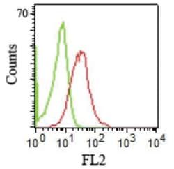
Antigen: Integrin alpha L/CD11a
Applications: Flow Cytometry, Immunocytochemistry, Immunofluorescence, Immunohistochemistry (Frozen), SDS-Page
Conjugate: Unconjugated
Host Species: Mouse
Research Discipline: Adaptive Immunity, Cellular Markers, Cytokine Research, Hematopoietic Stem Cell Markers, Immunology, Myeloid Cell Markers, Signal Transduction, Stem Cell Markers
Formulation: PBS with 0.05% BSA. with 0.05% Sodium Azide
Gene Alias: CD11 antigen-like family member A, CD11a antigen, CD11Aantigen CD11A (p180), lymphocyte function-associated antigen 1, alphapolypeptide, integrin alpha-L, integrin gene promoter, integrin, alpha L (antigen CD11A (p180), lymphocyte function-associated antigen1; alpha polypeptide), Leukocyte adhesion glycoprotein LFA-1 alpha chain, Leukocyte function-associated molecule 1 alpha chain, LFA-1, LFA-1 alpha, LFA1A, LFA-1A, lymphocyte function-associated antigen 1
Gene Symbols: ITGAL
Isotype: IgG2b κ
Purification Method: Protein A purified
Test Specificity: Recognizes a protein of 180kDa, identified as CD11a (Leucocyte Workshop II; Code N202). CD11a complex with the 2 subunit of the integrin family, CD18, to form the cell surface heterodimer, LFA-1 or CD11a/C18 (aLbL). LFA-1 is expressed on all leukocytes including lymphocytes, monocytes, and granulocytes. It is involved in leukocyte adhesion to its ligands including intercellular adhesion molecule-1 (ICAM-1 or CD54), ICAM-2 (CD102), ICAM-3 (CD50) and Telencephalin (TLN) and play a role in most immune/inflammatory responses.
Dilution: Flow Cytometry 0.5-1ug/million cells, Immunocytochemistry/Immunofluorescence 0.5-1ug/ml, Immunohistochemistry-Frozen 0.5-1.0ug/ml, SDS-Page
Classification: Monoclonal
Form: Purified
Regulatory Status: RUO
Target Species: Human
Gene Accession No.: P20701
Gene ID (Entrez): 3683
Immunogen: Stimulated human leukocytes
Primary or Secondary: Primary
Content And Storage: Store at 4C.
Molecular Weight of Antigen: 180 kDa