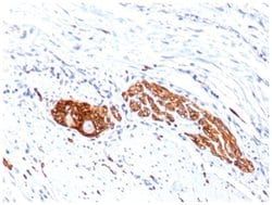
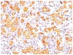
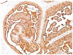
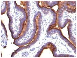
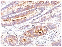
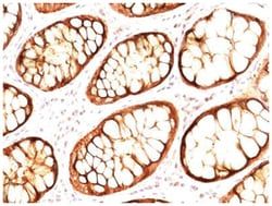
MUC1 Antibody (GP1.4 + E29), Novus Biologicals™
Mouse Monoclonal Antibody
Manufacturer: Fischer Scientific
The price for this product is unavailable. Please request a quote
Antigen
MUC-1
Dilution
Western Blot 0.5-1.0ug/ml, Flow Cytometry 0.5-1ug/million cells, Immunocytochemistry/Immunofluorescence 0.5-1ug/ml, Immunoprecipitation 0.5-1ug/500ug protein lysate, Immunohistochemistry-Paraffin 0.5-1ug/ml, Immunohistochemistry-Frozen 0.5-1ug/ml
Classification
Monoclonal
Form
Purified
Regulatory Status
RUO
Target Species
Human
Gene Accession No.
P15941
Gene ID (Entrez)
4582
Immunogen
Human milk fat globule membranes (GP1.4); Delipidated extract of human milk fat globule membranes (E29)
Primary or Secondary
Primary
Content And Storage
Store at 4C.
Clone
GP1.4 + E29
Applications
Western Blot, Flow Cytometry, Immunocytochemistry, Immunofluorescence, Immunoprecipitation, Immunohistochemistry (Paraffin)
Conjugate
Unconjugated
Host Species
Mouse
Research Discipline
Cancer, Cellular Markers, Extracellular Matrix
Formulation
PBS with 0.05% BSA. with 0.05% Sodium Azide
Gene Alias
Breast carcinoma-associated antigen DF3, Carcinoma-associated mucin, CD227, CD227 antigen, DF3 antigen, EMA, episialin, H23 antigen, H23AG, KL-6, MAM6, MUC-1, MUC1/ZD, mucin 1, cell surface associated, mucin 1, transmembrane, mucin-1, Peanut-reactive urinary mucin, PEMMUC-1/SEC, PEMT, Polymorphic epithelial mucin, PUMMUC-1/X, tumor associated epithelial mucin, Tumor-associated epithelial membrane antigen, Tumor-associated mucin
Gene Symbols
MUC1
Isotype
IgG1 κ, IgG2a κ
Purification Method
Protein A purified
Test Specificity
In Western blotting, it recognizes proteins in MW range of 265-400kDa, identified as different glycoforms of EMA. The alpha subunit has cell adhesive properties. It can act both as an adhesion and an anti-adhesion protein. EMA may provide a protective layer on epithelial cells against bacterial and enzyme attack. The beta subunit contains a C-terminal domain, which is involved in cell signaling, through phosphorylations and protein-protein interactions. In immunohistochemical assays, it superbly stains routine formalin/paraffin carcinoma tissues. Antibody to EMA is useful as a pan-epithelial marker for detecting early metastatic loci of carcinoma in bone marrow or liver.
Related Products


Description
- MUC1 Monoclonal specifically detects MUC1 in Human samples
- It is validated for Immunohistochemistry, Immunohistochemistry-Paraffin.
Compare Similar Items
Show Difference
Antigen: MUC-1
Dilution: Western Blot 0.5-1.0ug/ml, Flow Cytometry 0.5-1ug/million cells, Immunocytochemistry/Immunofluorescence 0.5-1ug/ml, Immunoprecipitation 0.5-1ug/500ug protein lysate, Immunohistochemistry-Paraffin 0.5-1ug/ml, Immunohistochemistry-Frozen 0.5-1ug/ml
Classification: Monoclonal
Form: Purified
Regulatory Status: RUO
Target Species: Human
Gene Accession No.: P15941
Gene ID (Entrez): 4582
Immunogen: Human milk fat globule membranes (GP1.4); Delipidated extract of human milk fat globule membranes (E29)
Primary or Secondary: Primary
Content And Storage: Store at 4C.
Clone: GP1.4 + E29
Applications: Western Blot, Flow Cytometry, Immunocytochemistry, Immunofluorescence, Immunoprecipitation, Immunohistochemistry (Paraffin)
Conjugate: Unconjugated
Host Species: Mouse
Research Discipline: Cancer, Cellular Markers, Extracellular Matrix
Formulation: PBS with 0.05% BSA. with 0.05% Sodium Azide
Gene Alias: Breast carcinoma-associated antigen DF3, Carcinoma-associated mucin, CD227, CD227 antigen, DF3 antigen, EMA, episialin, H23 antigen, H23AG, KL-6, MAM6, MUC-1, MUC1/ZD, mucin 1, cell surface associated, mucin 1, transmembrane, mucin-1, Peanut-reactive urinary mucin, PEMMUC-1/SEC, PEMT, Polymorphic epithelial mucin, PUMMUC-1/X, tumor associated epithelial mucin, Tumor-associated epithelial membrane antigen, Tumor-associated mucin
Gene Symbols: MUC1
Isotype: IgG1 κ, IgG2a κ
Purification Method: Protein A purified
Test Specificity: In Western blotting, it recognizes proteins in MW range of 265-400kDa, identified as different glycoforms of EMA. The alpha subunit has cell adhesive properties. It can act both as an adhesion and an anti-adhesion protein. EMA may provide a protective layer on epithelial cells against bacterial and enzyme attack. The beta subunit contains a C-terminal domain, which is involved in cell signaling, through phosphorylations and protein-protein interactions. In immunohistochemical assays, it superbly stains routine formalin/paraffin carcinoma tissues. Antibody to EMA is useful as a pan-epithelial marker for detecting early metastatic loci of carcinoma in bone marrow or liver.
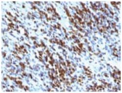
Antigen: Myogenin
Dilution: Western Blot 0.5-1.0ug/ml, Flow Cytometry 0.5-1ug/million cells, Immunocytochemistry/Immunofluorescence 0.5-1ug/ml, Immunoprecipitation 0.5-1ug/500ug protein lysate, Immunohistochemistry-Paraffin 0.5-1ug/ml, Immunohistochemistry-Frozen 0.5-1ug/ml
Classification: Monoclonal
Form: Purified
Regulatory Status: RUO
Target Species: Human, Mouse, Rat, Porcine, Feline
Gene Accession No.: P15173
Gene ID (Entrez): 4656
Immunogen: Human myogenin recombinant protein (MGN185); Rat myogenin recombinant fragment containing amino acid 30-224 (F5D)
Primary or Secondary: Primary
Content And Storage: Store at 4C.
Clone: MGN185 + F5D
Applications: Western Blot, Flow Cytometry, Immunocytochemistry, Immunofluorescence, Immunoprecipitation, Immunohistochemistry (Paraffin)
Conjugate: Unconjugated
Host Species: Mouse
Research Discipline: Cancer, Stem Cell Markers, Transcription Factors and Regulators
Formulation: PBS with 0.05% BSA. with 0.05% Sodium Azide
Gene Alias: BHLHC3, bHLHc3Myogenic factor-4; myogenin, Class C basic helix-loop-helix protein 3, myf-4, MYF4myogenin, Myogenic factor 4, MYOGENIN, myogenin (myogenic factor 4)
Gene Symbols: MYOG
Isotype: IgG
Purification Method: Protein A purified
Test Specificity: Myogenin is a member of the MyoD family of myogenic basic helix-loop-helix (bHLH) transcription factors that also includes MyoD, Myf-5, and MRF4 (also known as herculinor Myf-6). MyoD family members are expressed exclusively in skeletal muscle and play a key role in activating myogenesis by binding to enhancer sequences of muscle-specific genes. The regulatory domain of MyoD is approximately 70 amino acids in length and includes both a basic DNA binding motif and a bHLH dimerization motif. MyoD family members share about 80% amino acid homology in their bHLH motifs. Anti-myogenin labels the nuclei of myoblasts in developing muscle tissue, and is expressed in tumor cell nuclei of rhabdomyosarcoma and some leiomyosarcomas. Positive nuclear staining may occur in Wilms tumor.