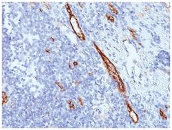
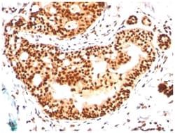
SUMO2/3 Antibody (SM23/496), Novus Biologicals™
Mouse Monoclonal Antibody
Manufacturer: Fischer Scientific
The price for this product is unavailable. Please request a quote
Antigen
SUMO2/3
Dilution
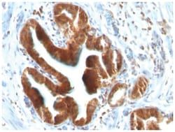
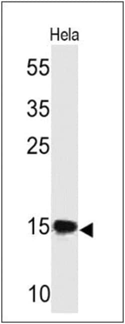
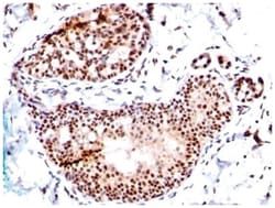
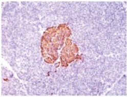
Western Blot 0.5-1ug/ml, Flow Cytometry 0.5-1ug/million cells, Immunocytochemistry/Immunofluorescence 0.5-1ug/ml, Immunoprecipitation 0.5-1ug/500ug protein lysate, Immunohistochemistry-Paraffin 0.5-1ug/ml, Immunohistochemistry-Frozen 0.5-1ug/ml
Classification
Monoclonal
Form
Purified
Regulatory Status
RUO
Formulation
PBS with 0.05% BSA. with 0.05% Sodium Azide
Gene ID (Entrez)
6613
Immunogen
Recombinant human SUMO2 protein
Primary or Secondary
Primary
Content And Storage
Store at 4C.
Clone
SM23/496
Applications
Western Blot, Flow Cytometry, Immunocytochemistry, Immunofluorescence, Immunoprecipitation, Immunohistochemistry (Paraffin)
Conjugate
Unconjugated
Host Species
Mouse
Target Species
Human
Gene Accession No.
P61956
Gene Symbols
SUMO2
Isotype
IgG1 κ
Purification Method
Protein A purified
Test Specificity
This MAb reacts with both SUMO-2 and SUMO-3. The small ubiquitin-related modifier (SUMO) proteins, which include SUMO-1, 2 and 3, belong to the ubiquitin-like protein family. Like ubiquitin, the SUMO proteins are synthesized as precursor proteins that undergo processing before conjugation to target proteins. Also, both utilize the E1, E2 and E3 cascade enzymes for conjugation. However, SUMO and ubiquitin differ with respect to targeting. Ubiquitination predominantly targets proteins for degradation, whereas sumoylation targets proteins to a variety of cellular processing, including nuclear transport, transcriptional regulation, apoptosis and protein stability. The unconjugated SUMO-1, 2 and 3 proteins localize to the nuclear membrane, nuclear bodies and cytoplasm, respectively. SUMO-1 utilizes Ubc9 for conjugation to several target proteins, which include MDM2, p53, PML and RanGap1. SUMO-2 and 3 contribute to a greater percentage of protein modification than does SUMO-1 and unlike SUMO
Related Products


Description
- SUMO2/3 Monoclonal specifically detects SUMO2/3 in Human samples
- It is validated for Western Blot, Flow Cytometry, Immunohistochemistry, Immunocytochemistry/Immunofluorescence, Immunohistochemistry-Paraffin, Flow (Intracellular).
Compare Similar Items
Show Difference
Antigen: SUMO2/3
Dilution: Western Blot 0.5-1ug/ml, Flow Cytometry 0.5-1ug/million cells, Immunocytochemistry/Immunofluorescence 0.5-1ug/ml, Immunoprecipitation 0.5-1ug/500ug protein lysate, Immunohistochemistry-Paraffin 0.5-1ug/ml, Immunohistochemistry-Frozen 0.5-1ug/ml
Classification: Monoclonal
Form: Purified
Regulatory Status: RUO
Formulation: PBS with 0.05% BSA. with 0.05% Sodium Azide
Gene ID (Entrez): 6613
Immunogen: Recombinant human SUMO2 protein
Primary or Secondary: Primary
Content And Storage: Store at 4C.
Clone: SM23/496
Applications: Western Blot, Flow Cytometry, Immunocytochemistry, Immunofluorescence, Immunoprecipitation, Immunohistochemistry (Paraffin)
Conjugate: Unconjugated
Host Species: Mouse
Target Species: Human
Gene Accession No.: P61956
Gene Symbols: SUMO2
Isotype: IgG1 κ
Purification Method: Protein A purified
Test Specificity: This MAb reacts with both SUMO-2 and SUMO-3. The small ubiquitin-related modifier (SUMO) proteins, which include SUMO-1, 2 and 3, belong to the ubiquitin-like protein family. Like ubiquitin, the SUMO proteins are synthesized as precursor proteins that undergo processing before conjugation to target proteins. Also, both utilize the E1, E2 and E3 cascade enzymes for conjugation. However, SUMO and ubiquitin differ with respect to targeting. Ubiquitination predominantly targets proteins for degradation, whereas sumoylation targets proteins to a variety of cellular processing, including nuclear transport, transcriptional regulation, apoptosis and protein stability. The unconjugated SUMO-1, 2 and 3 proteins localize to the nuclear membrane, nuclear bodies and cytoplasm, respectively. SUMO-1 utilizes Ubc9 for conjugation to several target proteins, which include MDM2, p53, PML and RanGap1. SUMO-2 and 3 contribute to a greater percentage of protein modification than does SUMO-1 and unlike SUMO
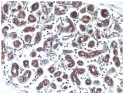
Antigen: TFF1/pS2
Dilution: Western Blot 0.5-1.0ug/ml, Flow Cytometry 0.5-1ug/million cells, Immunocytochemistry/Immunofluorescence 0.5-1ug/ml, Immunoprecipitation 0.5-1ug/500ug protein lysate, Immunohistochemistry-Paraffin 0.5-1ug/ml, Immunohistochemistry-Frozen 0.5-1ug/ml, SDS-Page
Classification: Monoclonal
Form: Purified
Regulatory Status: RUO
Formulation: PBS with 0.05% BSA. with 0.05% Sodium Azide
Gene ID (Entrez): 7031
Immunogen: Synthetic peptide of 28 amino acid residues corresponding to CFDDTVRGVPWCFYPNTIDVPPEEECEF (aa57-84) from the C-terminus of human pS2.
Primary or Secondary: Primary
Content And Storage: Store at 4C.
Clone: GE2 (same as R47/94)
Applications: Western Blot, Flow Cytometry, Immunocytochemistry, Immunofluorescence, Immunoprecipitation, Immunohistochemistry (Paraffin)
Conjugate: Unconjugated
Host Species: Mouse
Target Species: Human, Primate
Gene Accession No.: P04155
Gene Symbols: TFF1
Isotype: IgG1 κ
Purification Method: Protein A purified
Test Specificity: It recognizes a polypeptide of 6.5kDa, identified as pS2 estrogen-regulated protein. Its epitope is localized between aa57-84 of human pS2 protein. pS2 is a trefoil peptide. Trefoil peptides are protease resistant molecules secreted throughout the gut that play a role in mucosal healing. These peptides contain three intra-chain disulfide bonds, forming the trefoil motif, or P-domain. pS2 is known to form dimers and this dimerization is thought to play a role in its protective and healing properties. About 60% of breast carcinomas are positive for pS2. Staining is cytoplasmic, often with localization to the Golgi apparatus. pS2 is shown to be localized in normal stomach mucosa, gastric fluid, goblet cells in the colon and small intestine, and in ulcerations of the gastrointestinal tract. Several studies have shown that pS2 is primarily expressed in estrogen receptor-positive breast tumors and it may define a subset of estrogen-dependent tumors that displays an increased likelihood of re
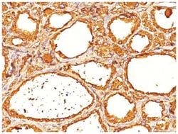
Antigen: Thyroglobulin
Dilution: Western Blot 0.5-1.0ug/ml, Flow Cytometry 0.5-1.0ug/million cells, Immunohistochemistry, Immunohistochemistry-Paraffin 1:100
Classification: Monoclonal
Form: Purified
Regulatory Status: RUO
Formulation: PBS with 0.05% BSA. with 0.05% Sodium Azide
Gene ID (Entrez): 7038
Immunogen: Human thyroid follicular cells
Primary or Secondary: Primary
Content And Storage: Store at 4C.
Clone: 2H11+6E1
Applications: Western Blot, Flow Cytometry, Immunohistochemistry, Immunohistochemistry (Paraffin)
Conjugate: Unconjugated
Host Species: Mouse
Target Species: Human, Mouse, Rat
Gene Accession No.: P01266
Gene Symbols: TG
Isotype: IgG
Purification Method: Protein A purified
Test Specificity: Thyroglobulin is a 660kDa dimeric pre-protein with mutiple glycosylation sites. It is produced by and processed within the thyroid gland to produce the hormone thyroxine and triiodothyronine. Prior to forming dimers, thyroglobulin monomers undergo conformational maturation in the endoplasmic reticulation. The vast majority of follicular carcinomas of the thyroid will give positive immunoreactivity for anti-thyroglobulin even though sometimes only focally. Poorly differentiated carcinomas of the thyroid are frequently anti-thyroglobulin negative. Adenocarcinomas of other-than-thyroid origin do not react with this antibody. This antibody is useful in identification of thyroid carcinoma of the papillary and follicular types. Presence of thyroglobulin in metastatic lesions establishes the thyroid origin of tumor. Anti-thyroglobulin, combined with anti-calcitonin, can identify medullary carcinomas of the thyroid. Furthermore, anti-thyroglobulin, combined with anti-TTF1, can be a reliable ma