PD-1 Antibody (SPM597) - Azide and BSA Free, Novus Biologicals™
Mouse Monoclonal Antibody
Manufacturer: Fischer Scientific
The price for this product is unavailable. Please request a quote
Antigen
PD-1
Concentration
1.0 mg/mL
Applications
Flow Cytometry, Immunohistochemistry (Paraffin), Immunofluorescence, CyTOF
Conjugate
Unconjugated
Host Species
Mouse
Research Discipline
Apoptosis, Stem Cell Markers
Formulation
PBS with No Preservative
Gene ID (Entrez)
5133
Immunogen
Recombinant full-length human PDCD1 protein
Primary or Secondary
Primary
Content And Storage
Store at 4C short term. Aliquot and store at -20C long term. Avoid freeze-thaw cycles.
Molecular Weight of Antigen
55 kDa
Clone
SPM597
Dilution
Flow Cytometry : 0.5 - 1 ug/million cells in 0.1 ml, Immunohistochemistry-Paraffin : 0.5 - 1.0 ug/ml, Immunofluorescence : 1 - 2 ug/ml, CyTOF-ready
Classification
Monoclonal
Form
Purified
Regulatory Status
RUO
Target Species
Human
Gene Alias
CD279, CD279 antigen, hPD-1, PD1hPD-l, programmed cell death 1, programmed cell death protein 1, Protein PD-1, SLEB2
Gene Symbols
PDCD1
Isotype
IgG1 κ
Purification Method
Protein A or G purified
Test Specificity
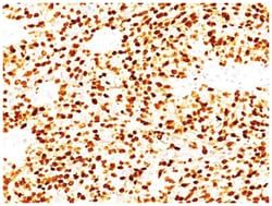
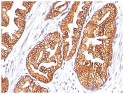
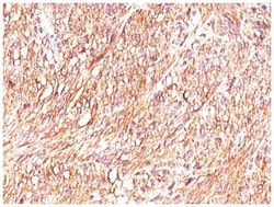
PDCD-1 (programmed cell death-1 protein), also designated CD279, is a type I transmembrane receptor and a member of the immunoglobin gene superfamily. It is expressed on activated T-cells, B-cells, and myeloid cells. Anti-PDCD-1 is a marker of angioimmunoblastic lymphoma and suggests a unique cell of origin for this neoplasm. Unlike CD10 and BCL6, PDCD-1 is expressed by few B-cells, so anti-PDCD-1 may be a more specific and useful diagnostic marker in angioimmunoblastic lymphoma. In addition, PDCD-1 expression provides evidence that angioimmunoblastic lymphoma is a neoplasm derived from germinal center-associated T-cells.
Related Products

Description
- PD-1 Monoclonal specifically detects PD-1 in Human samples
- It is validated for Flow Cytometry, Immunohistochemistry, Immunocytochemistry/Immunofluorescence, Immunohistochemistry-Paraffin, Immunofluorescence, CyTOF-ready.
Compare Similar Items
Show Difference
Antigen: PD-1
Concentration: 1.0 mg/mL
Applications: Flow Cytometry, Immunohistochemistry (Paraffin), Immunofluorescence, CyTOF
Conjugate: Unconjugated
Host Species: Mouse
Research Discipline: Apoptosis, Stem Cell Markers
Formulation: PBS with No Preservative
Gene ID (Entrez): 5133
Immunogen: Recombinant full-length human PDCD1 protein
Primary or Secondary: Primary
Content And Storage: Store at 4C short term. Aliquot and store at -20C long term. Avoid freeze-thaw cycles.
Molecular Weight of Antigen: 55 kDa
Clone: SPM597
Dilution: Flow Cytometry : 0.5 - 1 ug/million cells in 0.1 ml, Immunohistochemistry-Paraffin : 0.5 - 1.0 ug/ml, Immunofluorescence : 1 - 2 ug/ml, CyTOF-ready
Classification: Monoclonal
Form: Purified
Regulatory Status: RUO
Target Species: Human
Gene Alias: CD279, CD279 antigen, hPD-1, PD1hPD-l, programmed cell death 1, programmed cell death protein 1, Protein PD-1, SLEB2
Gene Symbols: PDCD1
Isotype: IgG1 κ
Purification Method: Protein A or G purified
Test Specificity: PDCD-1 (programmed cell death-1 protein), also designated CD279, is a type I transmembrane receptor and a member of the immunoglobin gene superfamily. It is expressed on activated T-cells, B-cells, and myeloid cells. Anti-PDCD-1 is a marker of angioimmunoblastic lymphoma and suggests a unique cell of origin for this neoplasm. Unlike CD10 and BCL6, PDCD-1 is expressed by few B-cells, so anti-PDCD-1 may be a more specific and useful diagnostic marker in angioimmunoblastic lymphoma. In addition, PDCD-1 expression provides evidence that angioimmunoblastic lymphoma is a neoplasm derived from germinal center-associated T-cells.
Antigen: MVP
Concentration: 1.0 mg/mL
Applications: Flow Cytometry, Immunohistochemistry (Paraffin), Immunofluorescence, CyTOF
Conjugate: Unconjugated
Host Species: Mouse
Research Discipline: Cancer
Formulation: PBS with No Preservative
Gene ID (Entrez): 9961
Immunogen: Proteins precipitated from human breast cancer MCF-7 cells
Primary or Secondary: Primary
Content And Storage: Store at 4C short term. Aliquot and store at -20C long term. Avoid freeze-thaw cycles.
Molecular Weight of Antigen: __
Clone: 1032
Dilution: Flow Cytometry : 0.5 - 1 ug/million cells in 0.1 ml, Immunohistochemistry-Paraffin : 0.5 - 1.0 ug/ml, Immunofluorescence : 0.5 - 1.0 ug/ml, CyTOF-ready
Classification: Monoclonal
Form: Purified
Regulatory Status: RUO
Target Species: Human
Gene Alias: LRPVAULT1, Lung resistance-related protein, major vault protein
Gene Symbols: MVP
Isotype: IgG1 κ
Purification Method: Protein A or G purified
Test Specificity: __
Antigen: TRIM29
Concentration: 1.0 mg/mL
Applications: Flow Cytometry, Immunohistochemistry (Paraffin), Immunofluorescence, CyTOF
Conjugate: Unconjugated
Host Species: Mouse
Research Discipline: __
Formulation: PBS with No Preservative
Gene ID (Entrez): 23650
Immunogen: Recombinant fragment (126 Amino acid residues between aa 1-200) of human TRIM29 protein
Primary or Secondary: Primary
Content And Storage: Store at 4C short term. Aliquot and store at -20C long term. Avoid freeze-thaw cycles.
Molecular Weight of Antigen: 66 kDa
Clone: TRIM29/1041
Dilution: Flow Cytometry : 0.5 - 1 ug/million cells in 0.1 ml, Immunohistochemistry-Paraffin : 0.5 - 1.0 ug/ml, Immunofluorescence : 0.5 - 1.0 ug/ml, CyTOF-ready
Classification: Monoclonal
Form: Purified
Regulatory Status: RUO
Target Species: Human
Gene Alias: Ataxia telangiectasia group D-associated protein, ATDCataxia-telangiectasia group D-associated protein, FLJ36085, tripartite motif containing 29, tripartite motif protein TRIM29, tripartite motif-containing 29, tripartite motif-containing protein 29
Gene Symbols: TRIM29
Isotype: IgG2a κ
Purification Method: Protein A or G purified
Test Specificity: It recognizes a 66kDa protein, which is identified as Tripartite motif-containing protein 29 (TRIM29). It interacts with the intermediate filament protein vimentin, a substrate for the PKC family of protein kinases, and with hPKCI-1, an inhibitor of the PKCs. TRIM29 protein contains both zinc finger and leucine zipper motifs, suggesting that the it may form homodimers and possibly associate with DNA. High expression of TRIM29 has been reported in gastric cancer and pancreatic cancer, and correlates with enhanced tumor growth and lymph node metastasis. TRIM29 is also able to distinguish lung squamous cell carcinoma from lung adenocarcinoma with ∼90% positive accuracy, when used in a panel with TTF-1, p63, CK5/6, and Napsin-A antibodies.