EpCAM/TROP1 Antibody (EGP40/826) - Azide and BSA Free, Novus Biologicals™
Mouse Monoclonal Antibody
Manufacturer: Fischer Scientific
The price for this product is unavailable. Please request a quote
Antigen
EpCAM/TROP1
Concentration
1.0 mg/mL
Applications
Western Blot, Flow Cytometry, Immunohistochemistry (Paraffin), Immunofluorescence, CyTOF
Conjugate
Unconjugated
Host Species
Mouse
Research Discipline
Cancer, Cancer Stem Cells
Formulation
PBS with No Preservative
Gene ID (Entrez)
4072
Immunogen
A synthetic peptide (around aa 20-60) from the N-terminus of human TACSTD1 protein.
Primary or Secondary
Primary
Content And Storage
Store at 4C short term. Aliquot and store at -20C long term. Avoid freeze-thaw cycles.
Molecular Weight of Antigen
41 kDa
Clone
EGP40/826
Dilution
Western Blot : 0.5 - 1.0 ug/ml, Flow Cytometry : 0.5 - 1 ug/million cells in 0.1 ml, Immunohistochemistry-Paraffin : 0.5 - 1.0 ug/ml, Immunofluorescence : 1 - 2 ug/ml, CyTOF-ready
Classification
Monoclonal
Form
Purified
Regulatory Status
RUO
Target Species
Human, Mouse (Negative), Rat (Negative)
Gene Alias
17-1A, 323/A3, ACSTD1, antigen identified by monoclonal AUA1, CD326 antigen, Cell surface glycoprotein Trop-1, chromosome 4, surface marker (35kD glycoprotein), DIAR5, EGP, EGP-2, EGP314, EGP40, EpCAM, epithelial cell adhesion molecule, Epithelial cell surface antigen, Epithelial glycoprotein, Epithelial glycoprotein 314, ESA, GA733-2EGP34, hEGP314, HNPCC8, KS 1/4 antigen, KS1/4, KSAHEA125, M1S2, M4S1Ly74, Major gastrointestinal tumor-associated protein GA733-2, MIC18MH99, MOC31, TACST-1, TACSTD1, TROP1CD326, Tumor-associated calcium signal transducer 1CO-17A
Gene Symbols
EPCAM
Isotype
IgG1 κ
Purification Method
Protein A or G purified
Test Specificity
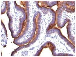
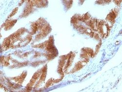
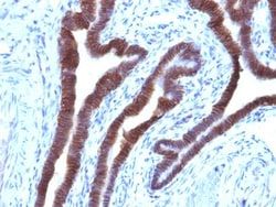
Recognizes a 40-43kDa transmembrane epithelial glycoprotein, identified as epithelial specific antigen (ESA), or epithelial cellular adhesion molecule (Ep-CAM). Ep-CAM is expressed on baso-lateral cell surface in most simple epithelia and a vast majority of carcinomas. This antibody has been used to distinguish adenocarcinoma from pleural mesothelioma and hepatocellular carcinoma. It is also useful in distinguishing serous carcinomas of the ovary from mesothelioma. This epithelial antigen plays an important role as a tumor-cell marker in lymph nodes from patients with esophageal carcinoma otherwise classified as node-negative. Epithelial antigen has also been suggested as a discriminator between basal cell and baso-squamous carcinomas, and squamous cell carcinoma of the skin.
Related Products


Description
- EpCAM/TROP1 Monoclonal specifically detects EpCAM/TROP1 in Human, Mouse (Negative), Rat (Negative) samples
- It is validated for Western Blot, Flow Cytometry, Immunohistochemistry, Immunocytochemistry/Immunofluorescence, Immunohistochemistry-Paraffin, Immunofluorescence, CyTOF-ready.
Compare Similar Items
Show Difference
Antigen: EpCAM/TROP1
Concentration: 1.0 mg/mL
Applications: Western Blot, Flow Cytometry, Immunohistochemistry (Paraffin), Immunofluorescence, CyTOF
Conjugate: Unconjugated
Host Species: Mouse
Research Discipline: Cancer, Cancer Stem Cells
Formulation: PBS with No Preservative
Gene ID (Entrez): 4072
Immunogen: A synthetic peptide (around aa 20-60) from the N-terminus of human TACSTD1 protein.
Primary or Secondary: Primary
Content And Storage: Store at 4C short term. Aliquot and store at -20C long term. Avoid freeze-thaw cycles.
Molecular Weight of Antigen: 41 kDa
Clone: EGP40/826
Dilution: Western Blot : 0.5 - 1.0 ug/ml, Flow Cytometry : 0.5 - 1 ug/million cells in 0.1 ml, Immunohistochemistry-Paraffin : 0.5 - 1.0 ug/ml, Immunofluorescence : 1 - 2 ug/ml, CyTOF-ready
Classification: Monoclonal
Form: Purified
Regulatory Status: RUO
Target Species: Human, Mouse (Negative), Rat (Negative)
Gene Alias: 17-1A, 323/A3, ACSTD1, antigen identified by monoclonal AUA1, CD326 antigen, Cell surface glycoprotein Trop-1, chromosome 4, surface marker (35kD glycoprotein), DIAR5, EGP, EGP-2, EGP314, EGP40, EpCAM, epithelial cell adhesion molecule, Epithelial cell surface antigen, Epithelial glycoprotein, Epithelial glycoprotein 314, ESA, GA733-2EGP34, hEGP314, HNPCC8, KS 1/4 antigen, KS1/4, KSAHEA125, M1S2, M4S1Ly74, Major gastrointestinal tumor-associated protein GA733-2, MIC18MH99, MOC31, TACST-1, TACSTD1, TROP1CD326, Tumor-associated calcium signal transducer 1CO-17A
Gene Symbols: EPCAM
Isotype: IgG1 κ
Purification Method: Protein A or G purified
Test Specificity: Recognizes a 40-43kDa transmembrane epithelial glycoprotein, identified as epithelial specific antigen (ESA), or epithelial cellular adhesion molecule (Ep-CAM). Ep-CAM is expressed on baso-lateral cell surface in most simple epithelia and a vast majority of carcinomas. This antibody has been used to distinguish adenocarcinoma from pleural mesothelioma and hepatocellular carcinoma. It is also useful in distinguishing serous carcinomas of the ovary from mesothelioma. This epithelial antigen plays an important role as a tumor-cell marker in lymph nodes from patients with esophageal carcinoma otherwise classified as node-negative. Epithelial antigen has also been suggested as a discriminator between basal cell and baso-squamous carcinomas, and squamous cell carcinoma of the skin.
Antigen: EpCAM/TROP1
Concentration: 1.0 mg/mL
Applications: Western Blot, Flow Cytometry, Immunohistochemistry (Paraffin), Immunofluorescence, CyTOF
Conjugate: Unconjugated
Host Species: Mouse
Research Discipline: Cancer, Cancer Stem Cells
Formulation: PBS with No Preservative
Gene ID (Entrez): 4072
Immunogen: Recombinant human Ep-CAM protein (full-length and fragments)
Primary or Secondary: Primary
Content And Storage: Store at 4C short term. Aliquot and store at -20C long term. Avoid freeze-thaw cycles.
Molecular Weight of Antigen: 41 kDa
Clone: PAN-EpCAM (Cocktail)
Dilution: Western Blot : 0.5 - 1.0 ug/ml, Flow Cytometry : 0.5 - 1 ug/million cells in 0.1 ml, Immunohistochemistry-Paraffin : 0.5 - 1.0 ug/ml, Immunofluorescence : 1 - 2 ug/ml, CyTOF-ready
Classification: Monoclonal
Form: Purified
Regulatory Status: RUO
Target Species: Human
Gene Alias: 17-1A, 323/A3, ACSTD1, antigen identified by monoclonal AUA1, CD326 antigen, Cell surface glycoprotein Trop-1, chromosome 4, surface marker (35kD glycoprotein), DIAR5, EGP, EGP-2, EGP314, EGP40, EpCAM, epithelial cell adhesion molecule, Epithelial cell surface antigen, Epithelial glycoprotein, Epithelial glycoprotein 314, ESA, GA733-2EGP34, hEGP314, HNPCC8, KS 1/4 antigen, KS1/4, KSAHEA125, M1S2, M4S1Ly74, Major gastrointestinal tumor-associated protein GA733-2, MIC18MH99, MOC31, TACST-1, TACSTD1, TROP1CD326, Tumor-associated calcium signal transducer 1CO-17A
Gene Symbols: EPCAM
Isotype: IgG1 κ
Purification Method: Protein A or G purified
Test Specificity: It is a cocktail of four highly specific monoclonal antibodies (EGP40/826, EGP40/837, EGP40/1110, EGP40/1120) that recognize extracellular as well as intracellular domains of the epithelial cellular adhesion molecule (Ep-CAM). It is a 40-43kDa transmembrane epithelial glycoprotein, identified as epithelial specific antigen (ESA), or Ep-CAM. Ep-CAM is expressed on baso-lateral cell surface in most simple epithelia and a vast majority of carcinomas. This epithelial antigen plays an important role as a tumor-cell marker in lymph nodes from patients with esophageal carcinoma otherwise classified as node-negative. Epithelial antigen has also been suggested as a discriminator between basal cell and baso-squamous carcinomas, and squamous cell carcinoma of the skin.
Antigen: CD43/Sialophorin
Concentration: 1.0 mg/mL
Applications: Flow Cytometry, Immunohistochemistry (Paraffin), Immunofluorescence, CyTOF
Conjugate: Unconjugated
Host Species: Mouse
Research Discipline: B Cell Development and Differentiation Markers, Immunology
Formulation: PBS with No Preservative
Gene ID (Entrez): 6693
Immunogen: Recombinant human SPN protein
Primary or Secondary: Primary
Content And Storage: Store at 4C short term. Aliquot and store at -20C long term. Avoid freeze-thaw cycles.
Molecular Weight of Antigen: __
Clone: SPN/1094
Dilution: Flow Cytometry : 0.5 - 1 ug/million cells in 0.1 ml, Immunohistochemistry-Paraffin : 0.5 - 1.0 ug/ml, Immunofluorescence : 1 - 2 ug/ml, CyTOF-ready
Classification: Monoclonal
Form: Purified
Regulatory Status: RUO
Target Species: Human
Gene Alias: CD43 antigen, CD43), Galactoglycoprotein, GALGP, Leukocyte sialoglycoprotein, Sialophorin, sialophorin (gpL115, leukosialin, CD43)
Gene Symbols: SPN
Isotype: IgG1 κ
Purification Method: Protein A or G purified
Test Specificity: It recognizes a cell surface glycoprotein of 95/115/135kDa (depending upon the extent of glycosylation), identified as CD43. 70-90% of T-cell lymphomas and from 22-37% of B-cell lymphomas express CD43. No reactivity has been observed with reactive B-cells. So a B-lineage population that co-expresses CD43 is highly likely to be a malignant lymphoma, especially a low-grade lymphoma, rather than a reactive B-cell population. When CD43 antibody is used in combination with anti-CD20, effective immunophenotyping of the lymphomas in formalin-fixed tissues can be obtained. Co-staining of a lymphoid infiltrate with anti-CD20 and anti-CD43 argues against a reactive process and favors a diagnosis of lymphoma.