Cytokeratin 7 Antibody (KRT7/760 + OV-TL12/30) - Azide and BSA Free, Novus Biologicals™
Mouse Monoclonal Antibody
Manufacturer: Fischer Scientific
The price for this product is unavailable. Please request a quote
Antigen
Cytokeratin 7
Concentration
1.0 mg/mL
Applications
Flow Cytometry, Immunohistochemistry (Paraffin), Immunofluorescence, CyTOF
Conjugate
Unconjugated
Host Species
Mouse
Research Discipline
Cytoskeleton Markers
Formulation
PBS with No Preservative
Gene ID (Entrez)
3855
Immunogen
Recombinant full-length human KRT7 protein (KRT7/760); OTN 11, ovarian carcinoma cell line (OV-TL12/30)
Primary or Secondary
Primary
Content And Storage
Store at 4C short term. Aliquot and store at -20C long term. Avoid freeze-thaw cycles.
Molecular Weight of Antigen
55 kDa
Clone
KRT7/760 + OV-TL12/30
Dilution
Flow Cytometry : 0.5 - 1 ug/million cells in 0.1 ml, Immunohistochemistry-Paraffin : 1 - 2 ug/ml, Immunofluorescence : 0.5 - 1.0 ug/ml, CyTOF-ready
Classification
Monoclonal
Form
Purified
Regulatory Status
RUO
Target Species
Human, Rat
Gene Alias
CK7, CK-7, cytokeratin 7, Cytokeratin-7, K2C7, K7keratin, 55K type II cytoskeletal, keratin 7, keratin, type II cytoskeletal 7, keratin-7, MGC129731, MGC3625, Sarcolectin, SCLkeratin, simple epithelial type I, K7, type II mesothelial keratin K7, Type-II keratin Kb7
Gene Symbols
KRT7
Isotype
IgG
Purification Method
Protein A or G purified
Test Specificity
It recognizes an intermediate filament protein (IFP) of 55kDa, which is identified as cytokeratin 7. This MAb is highly specific to cytokeratin 7 and shows no cross-reaction with other IFPs. Cytokeratin 7 is a basic cytokeratin, which is found in most glandular and transitional epithelia but not in the stratified squamous epithelia. Keratin 7 is expressed in the epithelial cells of ovary, lung, and breast but not of colon, prostate, or gastrointestinal tract. This MAb is highly useful in distinguishing ovarian carcinomas (keratin 7+) from colon carcinomas (keratin 7-).
Description
- Cytokeratin 7 Monoclonal specifically detects Cytokeratin 7 in Human, Rat samples
- It is validated for Western Blot, Flow Cytometry, Immunohistochemistry, Immunocytochemistry/Immunofluorescence, Immunohistochemistry-Paraffin, Immunofluorescence, CyTOF-ready.
Compare Similar Items
Show Difference

Antigen: Cytokeratin 7
Concentration: 1.0 mg/mL
Applications: Flow Cytometry, Immunohistochemistry (Paraffin), Immunofluorescence, CyTOF
Conjugate: Unconjugated
Host Species: Mouse
Research Discipline: Cytoskeleton Markers
Formulation: PBS with No Preservative
Gene ID (Entrez): 3855
Immunogen: Recombinant full-length human KRT7 protein (KRT7/760); OTN 11, ovarian carcinoma cell line (OV-TL12/30)
Primary or Secondary: Primary
Content And Storage: Store at 4C short term. Aliquot and store at -20C long term. Avoid freeze-thaw cycles.
Molecular Weight of Antigen: 55 kDa
Clone: KRT7/760 + OV-TL12/30
Dilution: Flow Cytometry : 0.5 - 1 ug/million cells in 0.1 ml, Immunohistochemistry-Paraffin : 1 - 2 ug/ml, Immunofluorescence : 0.5 - 1.0 ug/ml, CyTOF-ready
Classification: Monoclonal
Form: Purified
Regulatory Status: RUO
Target Species: Human, Rat
Gene Alias: CK7, CK-7, cytokeratin 7, Cytokeratin-7, K2C7, K7keratin, 55K type II cytoskeletal, keratin 7, keratin, type II cytoskeletal 7, keratin-7, MGC129731, MGC3625, Sarcolectin, SCLkeratin, simple epithelial type I, K7, type II mesothelial keratin K7, Type-II keratin Kb7
Gene Symbols: KRT7
Isotype: IgG
Purification Method: Protein A or G purified
Test Specificity: It recognizes an intermediate filament protein (IFP) of 55kDa, which is identified as cytokeratin 7. This MAb is highly specific to cytokeratin 7 and shows no cross-reaction with other IFPs. Cytokeratin 7 is a basic cytokeratin, which is found in most glandular and transitional epithelia but not in the stratified squamous epithelia. Keratin 7 is expressed in the epithelial cells of ovary, lung, and breast but not of colon, prostate, or gastrointestinal tract. This MAb is highly useful in distinguishing ovarian carcinomas (keratin 7+) from colon carcinomas (keratin 7-).
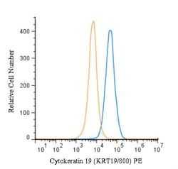
Antigen: Cytokeratin 19
Concentration: 1.0 mg/mL
Applications: Flow Cytometry, Immunohistochemistry (Paraffin), Immunofluorescence, CyTOF
Conjugate: Unconjugated
Host Species: Mouse
Research Discipline: Cancer, Cell Biology, Cellular Markers, Cytoskeleton Markers, Neuroscience, Stem Cell Markers
Formulation: PBS with No Preservative
Gene ID (Entrez): 3880
Immunogen: Recombinant human KRT19 protein
Primary or Secondary: Primary
Content And Storage: Store at 4C short term. Aliquot and store at -20C long term. Avoid freeze-thaw cycles.
Molecular Weight of Antigen: 40 kDa
Clone: KRT19/800
Dilution: Flow Cytometry : 0.5 - 1 ug/million cells in 0.1 ml, Immunohistochemistry-Paraffin : 0.5 - 1.0 ug/ml, Immunofluorescence : 1 - 2 ug/ml, CyTOF-ready
Classification: Monoclonal
Form: Purified
Regulatory Status: RUO
Target Species: Human, Mouse, Rat
Gene Alias: CK19, CK-19, cytokeratin 19, Cytokeratin-19,40-kDa keratin intermediate filament, K19cytokeratin-19, K1CS, keratin 19, keratin, type I cytoskeletal 19, keratin, type I, 40-kd, keratin-19, MGC15366
Gene Symbols: KRT19
Isotype: IgG1 κ
Purification Method: Protein A or G purified
Test Specificity: Recognizes a protein of 40kDa, identified as cytokeratin-19 (CK19), which is expressed in sweat gland, mammary gland ductal and secretory cells, bile ducts, gastrointestinal tract, bladder urothelium, oral epithelia, esophagus, and ectocervical epithelium. Anti-CK19 reacts with a wide variety of epithelial malignancies including adenocarcinomas of the colon, stomach, pancreas, biliary tract, liver, and breast. Perhaps the most useful application is the identification of thyroid carcinoma of the papillary type, although 50%-60% of follicular carcinomas are also labeled. Anti-CK19 is a useful marker for detection of tumor cells in lymph nodes, peripheral blood, bone marrow and breast cancer.
Antigen: Vimentin
Concentration: 1.0 mg/mL
Applications: Flow Cytometry, Immunohistochemistry (Paraffin), Immunofluorescence, CyTOF
Conjugate: Unconjugated
Host Species: Mouse
Research Discipline: Cancer, Cellular Markers, Cytoskeleton Markers, Growth and Development, Hypoxia, Neuronal Cell Markers, Neuronal Stem Cell Markers, Neuroscience, Signal Transduction, Stem Cell Markers, Stem Cells
Formulation: PBS with No Preservative
Gene ID (Entrez): 7431
Immunogen: Human thymic nuclear extract
Primary or Secondary: Primary
Content And Storage: Store at 4C short term. Aliquot and store at -20C long term. Avoid freeze-thaw cycles.
Molecular Weight of Antigen: __
Clone: LN-6
Dilution: Flow Cytometry : 0.5 - 1.0 ug/million cells in 0.1 ml, Immunohistochemistry-Paraffin : 1 - 2 ug/ml, Immunofluorescence : 0.5 - 1.0 ug/ml, CyTOF-ready
Classification: Monoclonal
Form: Purified
Regulatory Status: RUO
Target Species: Human, Mouse, Rat, Porcine, Feline, Primate, Rabbit, Sheep
Gene Alias: FLJ36605, vimentin
Gene Symbols: VIM
Isotype: IgM
Purification Method: Protein A or G purified
Test Specificity: This MAb reacts with a 58kDa protein identified as vimentin. It reacts with a non-hematopoietic epitope of vimentin and shows no cross-reaction with other closely related intermediate filament proteins (IFPs) such as desmin, keratin, neurofilament, and glial fibrillary acid protein.Vimentin is ubiquitously expressed in mesenchymal cells such as fibroblasts, smooth muscle cells, and endothelium. Antibody against vimentin is useful as part of an antibody panel for differential diagnosis of tumors of unknown origin.Ab-2 does not react with leukocyte common antigen-positive tissues such as lymphomas and leukemias.