Astrocytomas Antibody (J1-31), Novus Biologicals™
Mouse Monoclonal Antibody
Manufacturer: Fischer Scientific
The price for this product is unavailable. Please request a quote
Antigen
Astrocytomas
Dilution
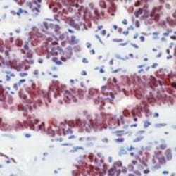
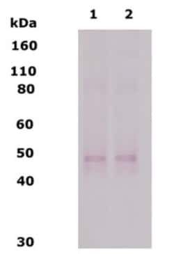
Western Blot 1:100-1:2000, Immunocytochemistry/Immunofluorescence 1:10-1:500, Immunohistochemistry-Paraffin 1:10-1:500
Classification
Monoclonal
Form
Ascites
Regulatory Status
RUO
Formulation
Ascites with No Preservative
Immunogen
Human cerebral white matter plaque materials from a multiple sclerosis patient.
Primary or Secondary
Primary
Content And Storage
Aliquot and store at -20C or -80C. Avoid freeze-thaw cycles.
Clone
J1-31
Applications
Immunocytochemistry, Western Blot, Immunohistochemistry (Paraffin), Immunofluorescence
Conjugate
Unconjugated
Host Species
Mouse
Target Species
Human, Rat
Gene Alias
Astrocytoma Marker, Astrocytomas Marker, Low Grade Astrocytoma Marker
Isotype
IgM
Purification Method
Unpurified
Test Specificity
The antibody recognizes an intracellular protein antigen (MW 30 kDa) expressed by human and rat astrocytes and other specialized glia (Muller cells of the retina, Bergmann fibers of the cerebellar cortex, tanycytes of the hypothalamus and ciliated ependymal cells) in the central nervous system (CNS).The antibody has recently been found to be a specific marker for low grade astrocytoma in human brain tissue. The antibody is able to distinguish between low grade astrocytoma and normal reactive gliosis (patent application filed). Monoclonal antibody J1-31 was raised against crude homogenate of brain tissue from a multiple sclerosis (MS) patient (autopsy sample; Malhotra et al.: Microbios Letters 26:151-157, 1984). In human brain, MAb J1-31 recognizes an intracellular protein antigen (J1-31 antigen), which bands at approximately 30,000 daltons under reducing conditions for sodium dodecyl sulfate gel electrophoresis (Singh et al.: Bioscience Reports 6:73-79, 1986). By immunofluorescence microscopy, MAb J1-31 stains those cells that are also stained by antiserum to glial fibrillary acidic protein (GFAP), namely astrocytes, retinal Muller cells, and tanycytes in the ependyma (Predy et al.: Bioscience Reports 7:491-502, 1987). In addition, MAb J1-31 stains ciliated ependymal cells that do not express GFAP (Malhotra, SK (1989) J Neurosci Res. 22(1):36-49).
Description
- Astrocytomas Monoclonal specifically detects Astrocytomas in Human, Rat samples
- It is validated for Western Blot, Immunohistochemistry, Immunocytochemistry/Immunofluorescence, Immunohistochemistry-Paraffin.
Compare Similar Items
Show Difference

Antigen: Astrocytomas
Dilution: Western Blot 1:100-1:2000, Immunocytochemistry/Immunofluorescence 1:10-1:500, Immunohistochemistry-Paraffin 1:10-1:500
Classification: Monoclonal
Form: Ascites
Regulatory Status: RUO
Formulation: Ascites with No Preservative
Immunogen: Human cerebral white matter plaque materials from a multiple sclerosis patient.
Primary or Secondary: Primary
Content And Storage: Aliquot and store at -20C or -80C. Avoid freeze-thaw cycles.
Clone: J1-31
Applications: Immunocytochemistry, Western Blot, Immunohistochemistry (Paraffin), Immunofluorescence
Conjugate: Unconjugated
Host Species: Mouse
Target Species: Human, Rat
Gene Alias: Astrocytoma Marker, Astrocytomas Marker, Low Grade Astrocytoma Marker
Isotype: IgM
Purification Method: Unpurified
Test Specificity: The antibody recognizes an intracellular protein antigen (MW 30 kDa) expressed by human and rat astrocytes and other specialized glia (Muller cells of the retina, Bergmann fibers of the cerebellar cortex, tanycytes of the hypothalamus and ciliated ependymal cells) in the central nervous system (CNS).The antibody has recently been found to be a specific marker for low grade astrocytoma in human brain tissue. The antibody is able to distinguish between low grade astrocytoma and normal reactive gliosis (patent application filed). Monoclonal antibody J1-31 was raised against crude homogenate of brain tissue from a multiple sclerosis (MS) patient (autopsy sample; Malhotra et al.: Microbios Letters 26:151-157, 1984). In human brain, MAb J1-31 recognizes an intracellular protein antigen (J1-31 antigen), which bands at approximately 30,000 daltons under reducing conditions for sodium dodecyl sulfate gel electrophoresis (Singh et al.: Bioscience Reports 6:73-79, 1986). By immunofluorescence microscopy, MAb J1-31 stains those cells that are also stained by antiserum to glial fibrillary acidic protein (GFAP), namely astrocytes, retinal Muller cells, and tanycytes in the ependyma (Predy et al.: Bioscience Reports 7:491-502, 1987). In addition, MAb J1-31 stains ciliated ependymal cells that do not express GFAP (Malhotra, SK (1989) J Neurosci Res. 22(1):36-49).
Antigen: MGMT
Dilution: __
Classification: Monoclonal
Form: __
Regulatory Status: __
Formulation: __
Immunogen: Recombinant human MGMT protein.
Primary or Secondary: Primary
Content And Storage: __
Clone: MT 3.1
Applications: Immunohistochemistry (Paraffin), Immunohistochemistry, Western Blot
Conjugate: Unconjugated
Host Species: Mouse
Target Species: Human
Gene Alias: 6-O-methylguanine-DNA methyltransferase, EC 2.1.1.63, methylated-DNA--protein-cysteine methyltransferase, methylguanine-DNA methyltransferase, O-6-methylguanine-DNA methyltransferase, O6-methylguanine-DNA methyltransferase, O-6-methylguanine-DNA-alkyltransferase
Isotype: IgG1
Purification Method: __
Test Specificity: This stains all mantle zone lymphocytes and 50% of germinal center lymphocytes. Basaloid epithelial cells of tonsil squamous mucosa were also stained positive with this.
Antigen: delta Opioid R/OPRD1
Dilution: Western Blot 1:100-1:2000
Classification: Polyclonal
Form: __
Regulatory Status: RUO
Formulation: PBS with 0.02% Sodium Azide
Immunogen: Synthetic peptide comprising residues 3-17 of the mouse and rat DOR-1 protein.
Primary or Secondary: Primary
Content And Storage: Store at 4C short term. Aliquot and store at -20C long term. Avoid freeze-thaw cycles.
Clone: __
Applications: Western Blot
Conjugate: Unconjugated
Host Species: Rabbit
Target Species: Mouse, Human, Rat
Gene Alias: delta opioid receptor 1, delta-type opioid receptor, D-OR-1, DOR-1, opioid receptor, delta 1, OPRD
Isotype: IgG
Purification Method: Affinity Purified
Test Specificity: Delta Opiod Receptor