Human EGFR (Research Grade Matuzumab Biosimilar) Antibody, R&D Systems™
Manufacturer: Fischer Scientific
Select a Size
| Pack Size | SKU | Availability | Price |
|---|---|---|---|
| Each of 1 | MAB10023100-Each-of-1 | In Stock | ₹ 46,858.50 |
MAB10023100 - Each of 1
In Stock
Quantity
1
Base Price: ₹ 46,858.50
GST (18%): ₹ 8,434.53
Total Price: ₹ 55,293.03
Antigen
EGFR
Classification
Monoclonal
Conjugate
Unconjugated
Formulation
Lyophilized from a 0.2 μm filtered solution in PBS with Trehalose. *Small pack size (SP) is supplied as a 0.2 μm filtered solution in PBS.
Host Species
Human
Purification Method
Protein A or G purified from cell culture supernatant
Primary or Secondary
Primary
Test Specificity
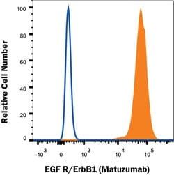
Detects human EGFR based on Matuzumab therapeutic antibody. This non-therapeutic antibody uses the same variable region sequence as the therapeutic antibody Matuzumab. This product is for research use only.
Target Species
Human
Form
Supernatant
Applications
Flow Cytometry, CyTOF
Clone
Hu104
Dilution
Flow Cytometry 0.25 μg/mL, CyTOF-ready
Gene Alias
avian erythroblastic leukemia viral (v-erb-b) oncogene homolog, cell growth inhibiting protein 40, cell proliferation-inducing protein 61, EC 2.7.10, EC 2.7.10.1, EGF R, epidermal growth factor receptor, epidermal growth factor receptor (avian erythroblastic leukemia viral (v-erb-b)oncogene homolog), ErbB, ErbB1, ERBB1PIG61, HER1, HER-1, mENA, Proto-oncogene c-ErbB-1, Receptor tyrosine-protein kinase erbB-1
Immunogen
Human EGFR,
Quantity
100 μg
Gene ID (Entrez)
1956.0
Reconstitution
Reconstitute at 0.5 mg/mL in sterile PBS.
Content And Storage
Use a manual defrost freezer and avoid repeated freeze-thaw cycles.12 months from date of receipt, -20 to -70 °C as supplied. 1 month, 2 to 8 °C under sterile conditions after reconstitution. 6 months, -20 to -70 °C under sterile conditions after reconstitution.
Isotype
IgG1
Related Products
Description
- EGFR Monoclonal specifically detects EGFR in Human samples
- It is validated for Flow Cytometry, CyTOF-ready.
Compare Similar Items
Show Difference
Antigen: EGFR
Classification: Monoclonal
Conjugate: Unconjugated
Formulation: Lyophilized from a 0.2 μm filtered solution in PBS with Trehalose. *Small pack size (SP) is supplied as a 0.2 μm filtered solution in PBS.
Host Species: Human
Purification Method: Protein A or G purified from cell culture supernatant
Primary or Secondary: Primary
Test Specificity: Detects human EGFR based on Matuzumab therapeutic antibody. This non-therapeutic antibody uses the same variable region sequence as the therapeutic antibody Matuzumab. This product is for research use only.
Target Species: Human
Form: Supernatant
Applications: Flow Cytometry, CyTOF
Clone: Hu104
Dilution: Flow Cytometry 0.25 μg/mL, CyTOF-ready
Gene Alias: avian erythroblastic leukemia viral (v-erb-b) oncogene homolog, cell growth inhibiting protein 40, cell proliferation-inducing protein 61, EC 2.7.10, EC 2.7.10.1, EGF R, epidermal growth factor receptor, epidermal growth factor receptor (avian erythroblastic leukemia viral (v-erb-b)oncogene homolog), ErbB, ErbB1, ERBB1PIG61, HER1, HER-1, mENA, Proto-oncogene c-ErbB-1, Receptor tyrosine-protein kinase erbB-1
Immunogen: Human EGFR,
Quantity: 100 μg
Gene ID (Entrez): 1956.0
Reconstitution: Reconstitute at 0.5 mg/mL in sterile PBS.
Content And Storage: Use a manual defrost freezer and avoid repeated freeze-thaw cycles.12 months from date of receipt, -20 to -70 °C as supplied. 1 month, 2 to 8 °C under sterile conditions after reconstitution. 6 months, -20 to -70 °C under sterile conditions after reconstitution.
Isotype: IgG1
Antigen: EGFR
Classification: Monoclonal
Conjugate: Unconjugated
Formulation: Lyophilized from a 0.2 μm filtered solution in PBS with Trehalose. *Small pack size (SP) is supplied as a 0.2 μm filtered solution in PBS.
Host Species: Human
Purification Method: Protein A or G purified from cell culture supernatant
Primary or Secondary: Primary
Test Specificity: Detects human EGFR based on Matuzumab therapeutic antibody. This non-therapeutic antibody uses the same variable region sequence as the therapeutic antibody Matuzumab. This product is for research use only.
Target Species: Human
Form: Lyophilized
Applications: Flow Cytometry, CyTOF
Clone: Hu104
Dilution: Flow Cytometry 0.25 μg/mL, CyTOF-ready
Gene Alias: avian erythroblastic leukemia viral (v-erb-b) oncogene homolog, cell growth inhibiting protein 40, cell proliferation-inducing protein 61, EC 2.7.10, EC 2.7.10.1, EGF R, epidermal growth factor receptor, epidermal growth factor receptor (avian erythroblastic leukemia viral (v-erb-b)oncogene homolog), ErbB, ErbB1, ERBB1PIG61, HER1, HER-1, mENA, Proto-oncogene c-ErbB-1, Receptor tyrosine-protein kinase erbB-1
Immunogen: Human EGFR,
Quantity: 25 μg
Gene ID (Entrez): 1956.0
Reconstitution: Reconstitute at 0.5 mg/mL in sterile PBS.
Content And Storage: Use a manual defrost freezer and avoid repeated freeze-thaw cycles.12 months from date of receipt, -20 to -70 °C as supplied. 1 month, 2 to 8 °C under sterile conditions after reconstitution. 6 months, -20 to -70 °C under sterile conditions after reconstitution.
Isotype: IgG1

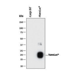
Antigen: Luciferase
Classification: Monoclonal
Conjugate: Unconjugated
Formulation: Lyophilized from a 0.2 μm filtered solution in PBS with Trehalose. *Small pack size (SP) is supplied as a 0.2 μm filtered solution in PBS.
Host Species: Mouse
Purification Method: Protein A or G purified from hybridoma culture supernatant
Primary or Secondary: Primary
Test Specificity: Detects NanoLuc™ (Nluc) Luciferase in direct ELISAs. Detects NanoLuc™ (Nluc) Luciferase and Large BiT in Western blots.
Target Species: Multi-species
Form: Lyophilized
Applications: Western Blot
Clone: 965808
Dilution: Western Blot 2 μg/mL
Gene Alias: ec 1.13.12.7, Fluc, luciferin 4 monooxygenase
Immunogen: Synthetic peptide of NanoLuc™ (Nluc) Luciferase
Quantity: 100 μg
Gene ID (Entrez): __
Reconstitution: Reconstitute at 0.5 mg/mL in sterile PBS.
Content And Storage: Use a manual defrost freezer and avoid repeated freeze-thaw cycles.12 months from date of receipt, -20 to -70°C as supplied. 1 month, 2 to 8°C under sterile conditions after reconstitution. 6 months, -20 to -70°C under sterile conditions after reconstitution.
Isotype: IgG2b