Human Bag-1 Antibody, R&D Systems™
Manufacturer: R&D Systems
Select a Size
| Pack Size | SKU | Availability | Price |
|---|---|---|---|
| Each of 1 | MAB852-Each-of-1 | In Stock | ₹ 42,225.16 |
MAB852 - Each of 1
In Stock
Quantity
1
Base Price: ₹ 42,225.16
GST (18%): ₹ 7,600.529
Total Price: ₹ 49,825.689
Antigen
Bag-1
Classification
Monoclonal
Concentration
LYOPH
Dilution
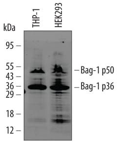
Western Blot 0.5 ug/mL, Immunoprecipitation 0.6 ug/10^6 cells, Knockout Validated
Gene Alias
BAG family molecular chaperone regulator 1, Bag1, BAG-1, Bcl-2 associating athanogene-1 protein, BCL2-associated athanogene, Bcl-2-associated athanogene 1, Bcl-2-binding protein, glucocortoid receptor-associated protein RAP46,46-KD, HAP, RAP46
Host Species
Mouse
Purification Method
Protein A or G purified from hybridoma culture supernatant
Regulatory Status
RUO
Gene ID (Entrez)
573
Reconstitution
Reconstitute at 0.5 mg/mL in sterile PBS.
Content And Storage
Use a manual defrost freezer and avoid repeated freeze-thaw cycles.12 months from date of receipt, -20 to -70 degreesC as supplied. 1 month, 2 to 8 degreesC under sterile conditions after reconstitution. 6 months, -20 to -70 degreesC under sterile conditions after reconstitution.
Isotype
IgG1
Applications
Western Blot, Immunoprecipitation, Immunoassay
Clone
GP3.10G3E2
Conjugate
Unconjugated
Formulation
Lyophilized from a 0.2 μm filtered solution in PBS with Trehalose. *Small pack size (SP) is supplied as a 0.2 μm filtered solution in PBS. with No Preservative
Gene Symbols
BAG1
Immunogen
E. coli-derived recombinant human Bag-1
Quantity
100 μg
Primary or Secondary
Primary
Test Specificity
Detects the 36ákDa and 50ákDa isoforms of human Bag-1. Does not detect mouse Bag-1.
Target Species
Human
Form
Purified
Related Products


Description
- Bag-1 Monoclonal specifically detects Bag-1 in Human samples
- It is validated for Western Blot, Immunoprecipitation, Knockout Validated.
Compare Similar Items
Show Difference
Antigen: Bag-1
Classification: Monoclonal
Concentration: LYOPH
Dilution: Western Blot 0.5 ug/mL, Immunoprecipitation 0.6 ug/10^6 cells, Knockout Validated
Gene Alias: BAG family molecular chaperone regulator 1, Bag1, BAG-1, Bcl-2 associating athanogene-1 protein, BCL2-associated athanogene, Bcl-2-associated athanogene 1, Bcl-2-binding protein, glucocortoid receptor-associated protein RAP46,46-KD, HAP, RAP46
Host Species: Mouse
Purification Method: Protein A or G purified from hybridoma culture supernatant
Regulatory Status: RUO
Gene ID (Entrez): 573
Reconstitution: Reconstitute at 0.5 mg/mL in sterile PBS.
Content And Storage: Use a manual defrost freezer and avoid repeated freeze-thaw cycles.12 months from date of receipt, -20 to -70 degreesC as supplied. 1 month, 2 to 8 degreesC under sterile conditions after reconstitution. 6 months, -20 to -70 degreesC under sterile conditions after reconstitution.
Isotype: IgG1
Applications: Western Blot, Immunoprecipitation, Immunoassay
Clone: GP3.10G3E2
Conjugate: Unconjugated
Formulation: Lyophilized from a 0.2 μm filtered solution in PBS with Trehalose. *Small pack size (SP) is supplied as a 0.2 μm filtered solution in PBS. with No Preservative
Gene Symbols: BAG1
Immunogen: E. coli-derived recombinant human Bag-1
Quantity: 100 μg
Primary or Secondary: Primary
Test Specificity: Detects the 36ákDa and 50ákDa isoforms of human Bag-1. Does not detect mouse Bag-1.
Target Species: Human
Form: Purified
Antigen: CD30/TNFRSF8
Classification: Monoclonal
Concentration: __
Dilution: Western Blot 1 ug/mL, ELISA Capture (Matched Antibody Pair) 2-8 ug/mL
Gene Alias: CD30, CD30 antigen, CD30KI-1, CD30L receptor, cytokine receptor CD30, D1S166EKi-1, Ki-1 antigen, Lymphocyte activation antigen CD30, TNFRSF8, tumor necrosis factor receptor superfamily member 8, tumor necrosis factor receptor superfamily, member 8
Host Species: Rat
Purification Method: Protein A or G purified from hybridoma culture supernatant
Regulatory Status: RUO
Gene ID (Entrez): 943
Reconstitution: Reconstitute at 0.5 mg/mL in sterile PBS.
Content And Storage: Use a manual defrost freezer and avoid repeated freeze-thaw cycles.12 months from date of receipt, -20 to -70 degreesC as supplied. 1 month, 2 to 8 degreesC under sterile conditions after reconstitution. 6 months, -20 to -70 degreesC under sterile conditions after reconstitution.
Isotype: IgG2a
Applications: Western Blot, ELISA
Clone: 115705
Conjugate: Unconjugated
Formulation: Lyophilized from a 0.2 μm filtered solution in PBS with Trehalose. *Small pack size (SP) is supplied as a 0.2 μm filtered solution in PBS. with No Preservative
Gene Symbols: TNFRSF8
Immunogen: Mouse myeloma cell line NS0-derived recombinant mouse CD30/TNFRSF8 Phe19-Thr281 Accession # Q60846
Quantity: 500 μg
Primary or Secondary: Primary
Test Specificity: Detects mouse CD30/TNFRSF8 in ELISAs and Western blots. In sandwich immunoassays, no cross-reactivity or interference with recombinant mouse (rm) TNF RI/TNFRSF1A, recombinant human (rh) TNF RI/TNFRSF1A, rhTNF RII/TNFRSF1B, rmTNF-alpha/TNFSF2, rhTNF-alpha/TNFSF2, rhTNF-beta/TNFSF1, rhCD30/TNFRSF8, or rmCD30 Ligand/TNFSF8 is observed. In Western blots, no cross-reactivity with rhNGFR/TNFRSF16, rmOPG/TNFRSF11B, rmRANK/TNFRSF11A, rmTNF RI/TNFRSF1A, rmTNF RII/TNFRSF1B, rmEDAR, rmTAJ/TNFSF19, rmLTR beta/TNFRSF3, rm4-1BB/TNFRSF9, rmCD27/TNFRSF7, rhCD30/TNFRSF8, rmCD40/TNFRSF5, rhDR3/TNFRSF12, rhDR6/TNFRSF21, rmFas/TNFRSF6, rmGITR/TNFRSF18, or rhHVEM/TNFRSF14 is observed.
Target Species: Mouse
Form: Purified
Antigen: CD30/TNFRSF8
Classification: Monoclonal
Concentration: __
Dilution: Western Blot 1 ug/mL, ELISA Capture (Matched Antibody Pair) 2-8 ug/mL
Gene Alias: CD30, CD30 antigen, CD30KI-1, CD30L receptor, cytokine receptor CD30, D1S166EKi-1, Ki-1 antigen, Lymphocyte activation antigen CD30, TNFRSF8, tumor necrosis factor receptor superfamily member 8, tumor necrosis factor receptor superfamily, member 8
Host Species: Rat
Purification Method: Protein A or G purified from hybridoma culture supernatant
Regulatory Status: RUO
Gene ID (Entrez): 943
Reconstitution: Reconstitute at 0.5 mg/mL in sterile PBS.
Content And Storage: Use a manual defrost freezer and avoid repeated freeze-thaw cycles.12 months from date of receipt, -20 to -70 degreesC as supplied. 1 month, 2 to 8 degreesC under sterile conditions after reconstitution. 6 months, -20 to -70 degreesC under sterile conditions after reconstitution.
Isotype: IgG2a
Applications: Western Blot, ELISA
Clone: 115705
Conjugate: Unconjugated
Formulation: Lyophilized from a 0.2 μm filtered solution in PBS with Trehalose. *Small pack size (SP) is supplied as a 0.2 μm filtered solution in PBS. with No Preservative
Gene Symbols: TNFRSF8
Immunogen: Mouse myeloma cell line NS0-derived recombinant mouse CD30/TNFRSF8 Phe19-Thr281 Accession # Q60846
Quantity: 25 μg
Primary or Secondary: Primary
Test Specificity: Detects mouse CD30/TNFRSF8 in ELISAs and Western blots. In sandwich immunoassays, no cross-reactivity or interference with recombinant mouse (rm) TNF RI/TNFRSF1A, recombinant human (rh) TNF RI/TNFRSF1A, rhTNF RII/TNFRSF1B, rmTNF-alpha/TNFSF2, rhTNF-alpha/TNFSF2, rhTNF-beta/TNFSF1, rhCD30/TNFRSF8, or rmCD30 Ligand/TNFSF8 is observed. In Western blots, no cross-reactivity with rhNGFR/TNFRSF16, rmOPG/TNFRSF11B, rmRANK/TNFRSF11A, rmTNF RI/TNFRSF1A, rmTNF RII/TNFRSF1B, rmEDAR, rmTAJ/TNFSF19, rmLTR beta/TNFRSF3, rm4-1BB/TNFRSF9, rmCD27/TNFRSF7, rhCD30/TNFRSF8, rmCD40/TNFRSF5, rhDR3/TNFRSF12, rhDR6/TNFRSF21, rmFas/TNFRSF6, rmGITR/TNFRSF18, or rhHVEM/TNFRSF14 is observed.
Target Species: Mouse
Form: Purified