Podoplanin, Mouse anti-Human, Clone: LpMab-17, Millipore Sigma™
Manufacturer: MilliporeSigma
Select a Size
| Pack Size | SKU | Availability | Price |
|---|---|---|---|
| Each of 1 | MABT86725UG-Each-of-1 | In Stock | ₹ 17,118.26 |
MABT86725UG - Each of 1
In Stock
Quantity
1
Base Price: ₹ 17,118.26
GST (18%): ₹ 3,081.287
Total Price: ₹ 20,199.547
Antigen
Podoplanin
Classification
Monoclonal
Conjugate
Unconjugated
Formulation
Purified mouse monoclonal antibody IgG1 in buffer containing 0.1 M Tris-Glycine (pH 7.4), 150 mM NaCl with 0.05% sodium azide.
Gene Symbols
PDPN;GP36;PSEC0003;PSEC0025
Immunogen
LN229 glioma cells expressing human Podoplanin.
Quantity
25 μg
Research Discipline
Cell Structure
Test Specificity
Clone LpMab-17 detects podoplanin in human cells. It detects PLAG4 domain. The minimal epitope seunce is shown to be Gly77-Asp82.
Content And Storage
Stable for 1 year at 2-8°C from date of receipt.
Isotype
IgG1 κ
Applications
ELISA, Flow Cytometry, Immunohistochemistry (Paraffin), Western Blot
Clone
LpMab-17
Dilution
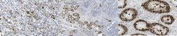
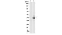
Immunohistochemistry Analysis: A 1:1,000 dilution from a representative lot detected Podoplanin in human testis cancer tissue. Flow Cytometry Analysis: A representative lot detected Podoplanin in Flow Cytometry applications (Kato, Y., et. al. (2016). Monoclon Antib Immunodiagn Immunother . 35(2):109-16). Immunohistochemistry Analysis: A representative lot detected Podoplanin in Immunohistochemistry applications (Kato, Y., et. al. (2016). Monoclon Antib Immunodiagn Immunother . 35(2):109-16). Western Blotting Analysis: A representative lot detected Podoplanin in Western Blotting applications (Kato, Y., et. al. (2016). Monoclon Antib Immunodiagn Immunother . 35(2):109-16). ELISA Analysis: A representative lot detected Podoplanin in ELISA applications (Kato, Y., et. al. (2016). Monoclon Antib Immunodiagn Immunother . 35(2):109-16).
Gene Alias
Aggrus;Glycoprotein 36;Gp36;PA2.26 antigen;T1-alpha;T1A
Host Species
Mouse
Purification Method
Protein G purified
Regulatory Status
RUO
Primary or Secondary
Primary
Target Species
Human
Form
Purified
Description
- Anti-Podoplanin, clone LpMab-17, Cat
- No
- MABT867, is a mouse monoclonal antibody that detects human podoplanin and has been tested for use in ELISA, Flow Cytometry, Immunohistochemistry (Paraffin), and Western Blotting
- Podoplanin (UniProt: Q86YL7; also known as Aggrus, Glycoprotein 36, Gp36, PA2.26 antigen, T1-alpha, T1A) is encoded by the PDPN (also known as GP36) gene (Gene ID: 10630) in human
- Podoplanin serves as the endogenous ligand of C-type lectin-like receptor-2 (CLEC-2) and is highly expressed in various tumors and in some normal cells, such as lymphatic endothelial cells and podocytes
- It may be involved in cell migration and/or actin cytoskeleton organization
- Podoplanin is localized to actin-rich microvilli and plasma membrane projections, such as filopodia, lamellipodia, and ruffles
- Six isoforms of podoplanin have been described that are produced by alternative splicing
- It is synthesized with a signal peptide of 22 amino acids, which is cleaved to produce the mature form
- Podoplanin contains an extracellular domain (aa 23-131), a helical domain (aa 132-152) and a cytoplasmic region (aa 153-162)
- Podoplanin possesses three tandem repeat of platelet aggregation-stimulating (PLAG) domains in its N-terminus
- Among the PLAG domains, sialylated O-glycan on Thr52 of PLAG3 is essential for the binding to C-type lectin-like receptor-2 (CLEC-2)
- Interaction of human podoplanin with CLEC-2 mainly involves Glu47 and Asp48 in the platelet PLAG3
- Clone LpMab-17 is reported to bind to non-PLAG domain and detects endogenous podoplanin in cancer cells and normal cells
- It recognizes glycan-deficient podoplanin indicating that its interaction with podoplanin is independent of its glycosylation
- The minimum epitope of LpMab-17 is identified as Gly77-Asp82
- Although human and monkey podoplanin exhibit 94% homology, this clone does not recognize monkey podoplanin
- (Ref.: Kato, Y., et al
- (2016)
- Monoclon
- Antib
- immunodiag
- Immunother
- 35(2); 109-116).
Compare Similar Items
Show Difference
Antigen: Podoplanin
Classification: Monoclonal
Conjugate: Unconjugated
Formulation: Purified mouse monoclonal antibody IgG1 in buffer containing 0.1 M Tris-Glycine (pH 7.4), 150 mM NaCl with 0.05% sodium azide.
Gene Symbols: PDPN;GP36;PSEC0003;PSEC0025
Immunogen: LN229 glioma cells expressing human Podoplanin.
Quantity: 25 μg
Research Discipline: Cell Structure
Test Specificity: Clone LpMab-17 detects podoplanin in human cells. It detects PLAG4 domain. The minimal epitope seunce is shown to be Gly77-Asp82.
Content And Storage: Stable for 1 year at 2-8°C from date of receipt.
Isotype: IgG1 κ
Applications: ELISA, Flow Cytometry, Immunohistochemistry (Paraffin), Western Blot
Clone: LpMab-17
Dilution: Immunohistochemistry Analysis: A 1:1,000 dilution from a representative lot detected Podoplanin in human testis cancer tissue. Flow Cytometry Analysis: A representative lot detected Podoplanin in Flow Cytometry applications (Kato, Y., et. al. (2016). Monoclon Antib Immunodiagn Immunother . 35(2):109-16). Immunohistochemistry Analysis: A representative lot detected Podoplanin in Immunohistochemistry applications (Kato, Y., et. al. (2016). Monoclon Antib Immunodiagn Immunother . 35(2):109-16). Western Blotting Analysis: A representative lot detected Podoplanin in Western Blotting applications (Kato, Y., et. al. (2016). Monoclon Antib Immunodiagn Immunother . 35(2):109-16). ELISA Analysis: A representative lot detected Podoplanin in ELISA applications (Kato, Y., et. al. (2016). Monoclon Antib Immunodiagn Immunother . 35(2):109-16).
Gene Alias: Aggrus;Glycoprotein 36;Gp36;PA2.26 antigen;T1-alpha;T1A
Host Species: Mouse
Purification Method: Protein G purified
Regulatory Status: RUO
Primary or Secondary: Primary
Target Species: Human
Form: Purified
Antigen: phospho-cytokeratin-18 (K18) (Ser33)
Classification: Monoclonal
Conjugate: Unconjugated
Formulation: Purified mouse monoclonal antibody IgG1 in buffer containing 0.1 M Tris-Glycine (pH 7.4), 150 mM NaCl with 0.05% sodium azide.
Gene Symbols: KRT18;CYK18;PIG46
Immunogen: KLH-conjugated linear peptide corresponding to 13 amino acids surrounding phosphorylated serine 33 (initiator methionine removed) from the N-terminal region of human cytokeratin-18.
Quantity: 100 μg
Research Discipline: Cell Structure
Test Specificity: Clone IB4 specifically detects human cytokeratin-18 phosphorylated on serine 33.
Content And Storage: Stable for 1 year at 2-8°C from date of receipt.
Isotype: IgG1 κ
Applications: Immunocytochemistry, Immunofluorescence, Immunoprecipitation, Western Blot
Clone: IB4
Dilution: Immunocytochemistry Analysis: A representative lot detected phospho-cytokeratin-18 (K18) (Ser33) in immunocytochemistry applications (Ku, N.O., et. al. (1998). EMBO J. 17(7):1892-906).Immunoprecipitation Analysis: A representative lot immunoprecipitated phospho-cytokeratin-18 (K18) (Ser33) in immunoprecipitation applications (Ku, N.O., et. al. (1998). EMBO J. 17(7):1892-906).Immunofluorescence Analysis: A representative lot detected phospho-cytokeratin-18 (K18) (Ser33) in Immunofluorescence applications (Ku, N.O., et. al. (1998). EMBO J. 17(7):1892-906).Western Blotting Analysis: A representative lot detected phospho-cytokeratin-18 (K18) (Ser33) in Western Blotting applications (Yoon, K.H., et. al. (2001). J Cell Biol. 153(3):503-16).
Gene Alias: Keratin type I cytoskeletal 18;Cell proliferation-inducing gene 46 protein;CK-18;Keratin-18;K18
Host Species: Mouse
Purification Method: Protein G purified
Regulatory Status: RUO
Primary or Secondary: Primary
Target Species: Human
Form: Purified
Antigen: phospho-cytokeratin-18 (K18) (Ser33)
Classification: Monoclonal
Conjugate: Unconjugated
Formulation: Purified mouse monoclonal antibody IgG1 in buffer containing 0.1 M Tris-Glycine (pH 7.4), 150 mM NaCl with 0.05% sodium azide.
Gene Symbols: KRT18;CYK18;PIG46
Immunogen: KLH-conjugated linear peptide corresponding to 13 amino acids surrounding phosphorylated serine 33 (initiator methionine removed) from the N-terminal region of human cytokeratin-18.
Quantity: 25 μg
Research Discipline: Cell Structure
Test Specificity: Clone IB4 specifically detects human cytokeratin-18 phosphorylated on serine 33.
Content And Storage: Stable for 1 year at 2-8°C from date of receipt.
Isotype: IgG1 κ
Applications: Immunocytochemistry, Immunofluorescence, Immunoprecipitation, Western Blot
Clone: IB4
Dilution: Immunocytochemistry Analysis: A representative lot detected phospho-cytokeratin-18 (K18) (Ser33) in immunocytochemistry applications (Ku, N.O., et. al. (1998). EMBO J. 17(7):1892-906).Immunoprecipitation Analysis: A representative lot immunoprecipitated phospho-cytokeratin-18 (K18) (Ser33) in immunoprecipitation applications (Ku, N.O., et. al. (1998). EMBO J. 17(7):1892-906).Immunofluorescence Analysis: A representative lot detected phospho-cytokeratin-18 (K18) (Ser33) in Immunofluorescence applications (Ku, N.O., et. al. (1998). EMBO J. 17(7):1892-906).Western Blotting Analysis: A representative lot detected phospho-cytokeratin-18 (K18) (Ser33) in Western Blotting applications (Yoon, K.H., et. al. (2001). J Cell Biol. 153(3):503-16).
Gene Alias: Keratin type I cytoskeletal 18;Cell proliferation-inducing gene 46 protein;CK-18;Keratin-18;K18
Host Species: Mouse
Purification Method: Protein G purified
Regulatory Status: RUO
Primary or Secondary: Primary
Target Species: Human
Form: Purified
