CD63 Antibody (LAMP3/2788) - Azide and BSA Free, Novus Biologicals™
Manufacturer: Novus Biologicals
Select a Size
| Pack Size | SKU | Availability | Price |
|---|---|---|---|
| Each of 1 | NB002965-Each-of-1 | In Stock | ₹ 58,562.00 |
NB002965 - Each of 1
In Stock
Quantity
1
Base Price: ₹ 58,562.00
GST (18%): ₹ 10,541.16
Total Price: ₹ 69,103.16
Antigen
CD63
Classification
Monoclonal
Conjugate
Unconjugated
Formulation
10mM PBS.
Gene Symbols
CD63
Immunogen
Recombinant human CD63 protein fragment (around aa100-197) (exact sequence is proprietary) (Uniprot: P08962 )
Quantity
100 μg
Primary or Secondary
Primary
Test Specificity
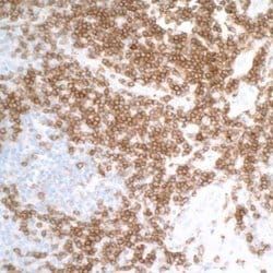
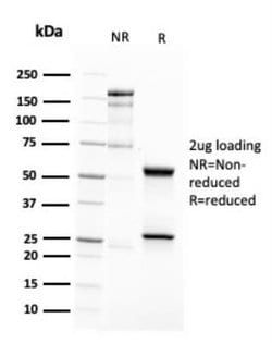
This monoclonal antibody recognizes protein of 26kDa-60kDa, which is identified as CD63. Its epitope is different from that of monoclonal antibody LAMP3/529. The tetraspanins are integral membrane proteins expressed on cell surface and granular membranes of hematopoietic cells and are components of multi-molecular complexes with specific integrins. The tetraspanin CD63 is a lysosomal membrane glycoprotein that translocates to the plasma membrane after platelet activation. CD63 is expressed on activated platelets, monocytes and macrophages, and is weakly expressed on granulocytes, T cell and B cells. It is located on the basophilic granule membranes and on the plasma membranes of lymphocytes and granulocytes. CD63 is a member of the TM4 superfamily of leukocyte glycoproteins that includes CD9, CD37 and CD53, which contain four transmembrane regions. CD63 may play a role in phagocytic and intracellular lysosome-phagosome fusion events. CD63 deficiency is associated with Hermansky-Pudlak
Content And Storage
Store at 4°C.
Isotype
IgG2b κ
Applications
Western Blot, Immunohistochemistry, Immunohistochemistry (Paraffin), Peptide Array
Clone
LAMP3/2788
Dilution
Western Blot 1-2 μg/mL, Immunohistochemistry 1-2 μg/mL, Immunohistochemistry-Paraffin 1-2 μg/mL, Protein Array
Gene Alias
CD63 antigen, CD63 antigen (melanoma 1 antigen), CD63 molecule, Granulophysin, LAMP-3, Lysosomal-associated membrane protein 3, ME491, melanoma 1 antigen, Melanoma-associated antigen ME491, melanoma-associated antigen MLA1, MLA1lysosome-associated membrane glycoprotein 3, Ocular melanoma-associated antigen, OMA81H, Tetraspanin-30, tspan-30, TSPAN30granulophysin
Host Species
Mouse
Purification Method
Protein A or G purified
Research Discipline
Autophagy, Cancer, Cardiovascular Biology, Cytokine Research, Immunology, Innate Immunity, Lysosome Markers, Metastasis
Gene ID (Entrez)
967
Target Species
Human, Mouse
Form
Purified
Related Products

Description
- CD63 Monoclonal specifically detects CD63 in Human, Mouse samples
- It is validated for Western Blot, Immunohistochemistry, Immunohistochemistry-Paraffin, Protein Array.
Compare Similar Items
Show Difference
Antigen: CD63
Classification: Monoclonal
Conjugate: Unconjugated
Formulation: 10mM PBS.
Gene Symbols: CD63
Immunogen: Recombinant human CD63 protein fragment (around aa100-197) (exact sequence is proprietary) (Uniprot: P08962 )
Quantity: 100 μg
Primary or Secondary: Primary
Test Specificity: This monoclonal antibody recognizes protein of 26kDa-60kDa, which is identified as CD63. Its epitope is different from that of monoclonal antibody LAMP3/529. The tetraspanins are integral membrane proteins expressed on cell surface and granular membranes of hematopoietic cells and are components of multi-molecular complexes with specific integrins. The tetraspanin CD63 is a lysosomal membrane glycoprotein that translocates to the plasma membrane after platelet activation. CD63 is expressed on activated platelets, monocytes and macrophages, and is weakly expressed on granulocytes, T cell and B cells. It is located on the basophilic granule membranes and on the plasma membranes of lymphocytes and granulocytes. CD63 is a member of the TM4 superfamily of leukocyte glycoproteins that includes CD9, CD37 and CD53, which contain four transmembrane regions. CD63 may play a role in phagocytic and intracellular lysosome-phagosome fusion events. CD63 deficiency is associated with Hermansky-Pudlak
Content And Storage: Store at 4°C.
Isotype: IgG2b κ
Applications: Western Blot, Immunohistochemistry, Immunohistochemistry (Paraffin), Peptide Array
Clone: LAMP3/2788
Dilution: Western Blot 1-2 μg/mL, Immunohistochemistry 1-2 μg/mL, Immunohistochemistry-Paraffin 1-2 μg/mL, Protein Array
Gene Alias: CD63 antigen, CD63 antigen (melanoma 1 antigen), CD63 molecule, Granulophysin, LAMP-3, Lysosomal-associated membrane protein 3, ME491, melanoma 1 antigen, Melanoma-associated antigen ME491, melanoma-associated antigen MLA1, MLA1lysosome-associated membrane glycoprotein 3, Ocular melanoma-associated antigen, OMA81H, Tetraspanin-30, tspan-30, TSPAN30granulophysin
Host Species: Mouse
Purification Method: Protein A or G purified
Research Discipline: Autophagy, Cancer, Cardiovascular Biology, Cytokine Research, Immunology, Innate Immunity, Lysosome Markers, Metastasis
Gene ID (Entrez): 967
Target Species: Human, Mouse
Form: Purified
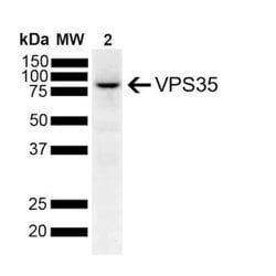
Antigen: VPS35
Classification: Monoclonal
Conjugate: Unconjugated
Formulation: PBS (pH 7.4), 50% glycerol with 0.09% Sodium Azide
Gene Symbols: VPS35
Immunogen: Full length recombinant human VSP35
Quantity: 100 μg
Primary or Secondary: Primary
Test Specificity: __
Content And Storage: Store at -20°C.
Isotype: __
Applications: Western Blot, Immunocytochemistry, Immunofluorescence, Immunoprecipitation, Immunoassay
Clone: 70000
Dilution: Western Blot 1:1000, Immunocytochemistry/Immunofluorescence 1:200, Immunoprecipitation 1:200, Knockout Validated
Gene Alias: DKFZp434P1672, FLJ10752, FLJ13588, FLJ20388, hVPS35, Maternal-embryonic 3, MEM3DKFZp434E1211, vacuolar protein sorting 35 (yeast homolog), vacuolar protein sorting 35 (yeast), vacuolar protein sorting 35 homolog (S. cerevisiae), vacuolar protein sorting-associated protein 35, Vesicle protein sorting 35
Host Species: Mouse
Purification Method: Protein G purified
Research Discipline: Autophagy, Cellular Markers
Gene ID (Entrez): 55737
Target Species: Human, Mouse, Rat
Form: Purified
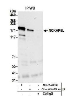
Antigen: NCKAP5L
Classification: Polyclonal
Conjugate: Unconjugated
Formulation: Tris-buffered Saline, 0.1% BSA with 0.09% Sodium Azide
Gene Symbols: NCKAP5L
Immunogen: The epitope recognized by A305-888A-M maps to a region between residue 1025 and 1075 of human Nck-associated protein 5-like using the numbering given in entry Q9HCH0.2 (GeneID 57701).
Quantity: 100 μL
Primary or Secondary: Primary
Test Specificity: __
Content And Storage: Store at 2-8°C/ 1 year from date of receipt
Isotype: __
Applications: Western Blot, Immunoprecipitation
Clone: __
Dilution: Western Blot 1:1000, Immunoprecipitation 50-100 μL/mg lysate
Gene Alias: FLJ36581, KIAA1602, NCK-associated protein 5-like
Host Species: Rabbit
Purification Method: Affinity Purified
Research Discipline: __
Gene ID (Entrez): 57701
Target Species: Human, Mouse
Form: __