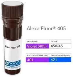
NB003496

Histone H2AX, p Ser139 Antibody, DyLight 680, Novus Biologicals™

Manufacturer: Novus Biologicals

Select a Size

Pack Size SKU Availability Price
Each of 1 NB003496-Each-of-1 In Stock ₹ 67,763.52

NB003496 - Each of 1

₹ 67,763.52

In Stock

Quantity

1

Base Price: ₹ 67,763.52

GST (18%): ₹ 12,197.434

Total Price: ₹ 79,960.954

Antigen

Histone H2AX (p Ser139)

Classification

Polyclonal

Dilution

Western Blot, Flow Cytometry, Immunohistochemistry, Immunocytochemistry/Immunofluorescence, Immunohistochemistry-Paraffin, Immunohistochemistry-Frozen, Knockout Validated

Gene Alias

H2A.X, H2A/X, H2AFX

Host Species

Rabbit

Purification Method

Affinity Purified

Regulatory Status

RUO

Primary or Secondary

Primary

Target Species

Human, Mouse, Rat, Canine

Isotype

IgG

Applications

Western Blot, Flow Cytometry, Immunohistochemistry, Immunocytochemistry, Immunofluorescence, Immunohistochemistry (Paraffin), Immunohistochemistry (Frozen), Immunoassay

Conjugate

DyLight 680

Formulation

50mM Sodium Borate with 0.05% Sodium Azide

Gene Symbols

H2AX

Immunogen

This Histone H2AX [p Ser139] Antibody was developed against to a region surrounding phosphorylated serine 139 of human histone H2AX [Swiss-Prot entry P16104] (GeneID 3014).

Quantity

0.1 mL

Research Discipline

Checkpoint signaling, DNA Double Strand Break Repair, DNA Repair, Epigenetics, Mitotic Regulators, Phospho Specific

Test Specificity

The epitope maps to a region surrounding phosphorylated serine 139 of human histone H2AX.

Content And Storage

Store at 4°C in the dark.

Related Products

Img

Novus Biologicals

NB003495

--

Img

Novus Biologicals

NB003497

--

Img

Novus Biologicals

NB002012

--

Img

Novus Biologicals

NB002354

--

Img

Novus Biologicals

NB002349

--

Img

Novus Biologicals

NB002334

--

Img

Novus Biologicals

NB002340

--

Img

Novus Biologicals

NB002351

--

Description

  • Description Histone H2AX (p Ser139) Polyclonal antibody specifically detects Histone H2AX (p Ser139) in Human, Mouse, Rat, Canine samples
  • It is validated for Western Blot, Flow Cytometry, Immunohistochemistry, Immunocytochemistry, Immunofluorescence, Immunohistochemistry (Paraffin), Immunohistochemistry (Frozen), Immunoassay.

Compare Similar Items

Show Difference

Img

Novus Biologicals

NB003496

--


Antigen:
Histone H2AX (p Ser139)

Classification:
Polyclonal

Dilution:
Western Blot, Flow Cytometry, Immunohistochemistry, Immunocytochemistry/Immunofluorescence, Immunohistochemistry-Paraffin, Immunohistochemistry-Frozen, Knockout Validated

Gene Alias:
H2A.X, H2A/X, H2AFX

Host Species:
Rabbit

Purification Method:
Affinity Purified

Regulatory Status:
RUO

Primary or Secondary:
Primary

Target Species:
Human, Mouse, Rat, Canine

Isotype:
IgG

Applications:
Western Blot, Flow Cytometry, Immunohistochemistry, Immunocytochemistry, Immunofluorescence, Immunohistochemistry (Paraffin), Immunohistochemistry (Frozen), Immunoassay

Conjugate:
DyLight 680

Formulation:
50mM Sodium Borate with 0.05% Sodium Azide

Gene Symbols:
H2AX

Immunogen:
This Histone H2AX [p Ser139] Antibody was developed against to a region surrounding phosphorylated serine 139 of human histone H2AX [Swiss-Prot entry P16104] (GeneID 3014).

Quantity:
0.1 mL

Research Discipline:
Checkpoint signaling, DNA Double Strand Break Repair, DNA Repair, Epigenetics, Mitotic Regulators, Phospho Specific

Test Specificity:
The epitope maps to a region surrounding phosphorylated serine 139 of human histone H2AX.

Content And Storage:
Store at 4°C in the dark.

Img

Novus Biologicals

NB003497

--


Antigen:
Histone H2AX (p Ser139)

Classification:
Polyclonal

Dilution:
Western Blot, Simple Western, Immunohistochemistry, Immunohistochemistry-Paraffin, Immunohistochemistry-Frozen, Knockout Validated

Gene Alias:
H2A.X, H2A/X, H2AFX

Host Species:
Rabbit

Purification Method:
Affinity Purified

Regulatory Status:
RUO

Primary or Secondary:
Primary

Target Species:
Human, Mouse, Rat, Canine

Isotype:
IgG

Applications:
Western Blot, Immunohistochemistry, Immunohistochemistry (Paraffin), Immunohistochemistry (Frozen), Immunoassay

Conjugate:
HRP

Formulation:
PBS with No Preservative

Gene Symbols:
H2AX

Immunogen:
This Histone H2AX [p Ser139] Antibody was developed against to a region surrounding phosphorylated serine 139 of human histone H2AX [Swiss-Prot entry P16104] (GeneID 3014).

Quantity:
0.1 mL

Research Discipline:
Checkpoint signaling, DNA Double Strand Break Repair, DNA Repair, Epigenetics, Mitotic Regulators, Phospho Specific

Test Specificity:
The epitope maps to a region surrounding phosphorylated serine 139 of human histone H2AX.

Content And Storage:
Store at 4°C in the dark.

Img

Novus Biologicals

NB003498

--


Antigen:
14-3-3 gamma (ac Val2)

Classification:
Monoclonal

Dilution:
Western Blot, Immunocytochemistry/Immunofluorescence

Gene Alias:
14-3-3 gamma, 14-3-3 protein gamma, 14-3-3GAMMA, KCIP-1, Protein kinase C inhibitor protein 1, tyrosine 3-monooxygenase/tryptophan 5-monooxygenase activation protein, gammapolypeptide

Host Species:
Mouse

Purification Method:
Protein G purified

Regulatory Status:
RUO

Primary or Secondary:
Primary

Target Species:
Human

Isotype:
IgG2a

Applications:
Western Blot, Immunocytochemistry, Immunofluorescence

Conjugate:
Alexa Fluor 350

Formulation:
50mM Sodium Borate with 0.05% Sodium Azide

Gene Symbols:
YWHAG

Immunogen:
N-terminal fragment of human 14-3-3 gamma where the N-terminal Val was acetylated [UniProt# P61981]

Quantity:
0.1 mL

Research Discipline:
Cancer, Cell Cycle and Replication, Cellular Markers, Growth and Development, Neuronal Cell Markers, Neuroscience, Signal Transduction, Stem Cell Markers

Test Specificity:
This antibody is specific for the naturally occurring human form of 14-3-3 gamma, where the N-terminal Met is removed, resulting in an acetylated N-terminal Val. Unprocessed (non-modified) 14-3-3 gamma is not recognized by this antibody.

Content And Storage:
Store at 4°C in the dark.

Img

Novus Biologicals

NB003499

--


Antigen:
14-3-3 gamma (ac Val2)

Classification:
Monoclonal

Dilution:
Western Blot, Immunocytochemistry/Immunofluorescence

Gene Alias:
14-3-3 gamma, 14-3-3 protein gamma, 14-3-3GAMMA, KCIP-1, Protein kinase C inhibitor protein 1, tyrosine 3-monooxygenase/tryptophan 5-monooxygenase activation protein, gammapolypeptide

Host Species:
Mouse

Purification Method:
Protein G purified

Regulatory Status:
RUO

Primary or Secondary:
Primary

Target Species:
Human

Isotype:
IgG2a

Applications:
Western Blot, Immunocytochemistry, Immunofluorescence

Conjugate:
Alexa Fluor 405

Formulation:
50mM Sodium Borate with 0.05% Sodium Azide

Gene Symbols:
YWHAG

Immunogen:
N-terminal fragment of human 14-3-3 gamma where the N-terminal Val was acetylated [UniProt# P61981]

Quantity:
0.1 mL

Research Discipline:
Cancer, Cell Cycle and Replication, Cellular Markers, Growth and Development, Neuronal Cell Markers, Neuroscience, Signal Transduction, Stem Cell Markers

Test Specificity:
This antibody is specific for the naturally occurring human form of 14-3-3 gamma, where the N-terminal Met is removed, resulting in an acetylated N-terminal Val. Unprocessed (non-modified) 14-3-3 gamma is not recognized by this antibody.

Content And Storage:
Store at 4°C in the dark.