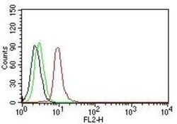
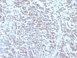
Cyclin D1 Antibody (DCS-6), FITC, Novus Biologicals™
Manufacturer: Novus Biologicals
Select a Size
| Pack Size | SKU | Availability | Price |
|---|---|---|---|
| Each of 1 | NB005687-Each-of-1 | In Stock | ₹ 57,494.00 |
NB005687 - Each of 1
In Stock
Quantity
1
Base Price: ₹ 57,494.00
GST (18%): ₹ 10,348.92
Total Price: ₹ 67,842.92
Antigen
Cyclin D1
Classification
Monoclonal
Conjugate
FITC
Formulation
PBS with 0.05% Sodium Azide
Gene Symbols
CCND1
Immunogen
Human full length recombinant cyclin D1 protein (Uniprot: P24385)
Quantity
0.1 mL
Research Discipline
Cancer, Cell Cycle and Replication, Core ESC Like Genes, mTOR Pathway, Stem Cell Markers, Wnt Signaling Pathway
Test Specificity
Recognizes a protein of 36kDa, identified as cyclin D1. Cyclin D1, one of the key cell cycle regulators, is a putative proto-oncogene overexpressed in a wide variety of human neoplasms. This antibody neutralizes the activity of cyclin D1 in vivo. About 60% of mantle cell lymphomas (MCL) contain a t(11; 14)(q13; q32) translocation resulting in over-expression of cyclin D1. This antibody is useful in identifying mantle cell lymphomas (cyclin D1 positive) from CLL/SLL and follicular lymphomas (cyclin D1 negative). About 40% of breast carcinomas are positive for Cyclin D1. Occasionally, hairy cell leukemia and plasma cell myeloma weakly express Cyclin D1.
Content And Storage
Store at 4°C in the dark.
Applications
Flow Cytometry, ELISA
Clone
DCS-6
Dilution
Flow Cytometry, ELISA
Gene Alias
B-cell lymphoma 1 protein, BCL-1, BCL-1 oncogene, BCL1D11S287E, cyclin D1, cyclin D1 (PRAD1: parathyroid adenomatosis 1), G1/S-specific cyclin D1, G1/S-specific cyclin-D1, PRAD1 oncogene, PRAD1B-cell CLL/lymphoma 1, U21B31
Host Species
Mouse
Purification Method
Protein A or G purified
Regulatory Status
RUO
Primary or Secondary
Primary
Target Species
Human, Mouse, Rat, Primate
Isotype
IgG2a κ
Related Products


Description
- Cyclin D1 Monoclonal specifically detects Cyclin D1 in Human, Mouse, Rat, Monkey samples
- It is validated for Flow Cytometry.
Compare Similar Items
Show Difference

Antigen: Cyclin D1
Classification: Monoclonal
Conjugate: FITC
Formulation: PBS with 0.05% Sodium Azide
Gene Symbols: CCND1
Immunogen: Human full length recombinant cyclin D1 protein (Uniprot: P24385)
Quantity: 0.1 mL
Research Discipline: Cancer, Cell Cycle and Replication, Core ESC Like Genes, mTOR Pathway, Stem Cell Markers, Wnt Signaling Pathway
Test Specificity: Recognizes a protein of 36kDa, identified as cyclin D1. Cyclin D1, one of the key cell cycle regulators, is a putative proto-oncogene overexpressed in a wide variety of human neoplasms. This antibody neutralizes the activity of cyclin D1 in vivo. About 60% of mantle cell lymphomas (MCL) contain a t(11; 14)(q13; q32) translocation resulting in over-expression of cyclin D1. This antibody is useful in identifying mantle cell lymphomas (cyclin D1 positive) from CLL/SLL and follicular lymphomas (cyclin D1 negative). About 40% of breast carcinomas are positive for Cyclin D1. Occasionally, hairy cell leukemia and plasma cell myeloma weakly express Cyclin D1.
Content And Storage: Store at 4°C in the dark.
Applications: Flow Cytometry, ELISA
Clone: DCS-6
Dilution: Flow Cytometry, ELISA
Gene Alias: B-cell lymphoma 1 protein, BCL-1, BCL-1 oncogene, BCL1D11S287E, cyclin D1, cyclin D1 (PRAD1: parathyroid adenomatosis 1), G1/S-specific cyclin D1, G1/S-specific cyclin-D1, PRAD1 oncogene, PRAD1B-cell CLL/lymphoma 1, U21B31
Host Species: Mouse
Purification Method: Protein A or G purified
Regulatory Status: RUO
Primary or Secondary: Primary
Target Species: Human, Mouse, Rat, Primate
Isotype: IgG2a κ

Antigen: CD43/Sialophorin
Classification: Monoclonal
Conjugate: Alexa Fluor 350
Formulation: 50mM Sodium Borate with 0.05% Sodium Azide
Gene Symbols: SPN
Immunogen: Myeloblastic KG1 cells were used as the immunogen for this antibody.
Quantity: 0.1 mL
Research Discipline: B Cell Development and Differentiation Markers, Immunology
Test Specificity: It recognizes a cell surface glycoprotein of 95/115/135kDa (depending upon the extent of glycosylation), identified as CD43 [Workshop IV]. Epitope of monoclonal antibody Bra7G is clearly different from that of monoclonal antibody DF-T1, called b as opposed to a for DF-T1. 70-90% of T-cell lymphomas and from 22-37% of B-cell lymphomas express CD43. No reactivity has been observed with reactive B-cells. So a B-lineage population that co-expresses CD43 is highly likely to be a malignant lymphoma, especially a low-grade lymphoma, rather than a reactive B-cell population. When CD43 antibody is used in combination with anti-CD20, effective immunophenotyping of the lymphomas in formalin-fixed tissues can be obtained. Co-staining of a lymphoid infiltrate with anti-CD20 and anti-CD43 argues against a reactive process and favors a diagnosis of lymphoma.
Content And Storage: Store at 4°C in the dark.
Applications: Western Blot, Flow Cytometry, ELISA, Immunocytochemistry, Immunofluorescence, Immunohistochemistry (Paraffin)
Clone: DF-T1
Dilution: Western Blot, Flow Cytometry, ELISA, Immunocytochemistry/Immunofluorescence, Immunohistochemistry-Paraffin
Gene Alias: CD43 antigen, CD43), Galactoglycoprotein, GALGP, Leukocyte sialoglycoprotein, Sialophorin, sialophorin (gpL115, leukosialin, CD43)
Host Species: Mouse
Purification Method: Protein A or G purified
Regulatory Status: RUO
Primary or Secondary: Primary
Target Species: Human
Isotype: IgG1 κ

Antigen: CD43/Sialophorin
Classification: Monoclonal
Conjugate: Alexa Fluor 532
Formulation: 50mM Sodium Borate with 0.05% Sodium Azide
Gene Symbols: SPN
Immunogen: Myeloblastic KG1 cells were used as the immunogen for this antibody.
Quantity: 0.1 mL
Research Discipline: B Cell Development and Differentiation Markers, Immunology
Test Specificity: It recognizes a cell surface glycoprotein of 95/115/135kDa (depending upon the extent of glycosylation), identified as CD43 [Workshop IV]. Epitope of monoclonal antibody Bra7G is clearly different from that of monoclonal antibody DF-T1, called b as opposed to a for DF-T1. 70-90% of T-cell lymphomas and from 22-37% of B-cell lymphomas express CD43. No reactivity has been observed with reactive B-cells. So a B-lineage population that co-expresses CD43 is highly likely to be a malignant lymphoma, especially a low-grade lymphoma, rather than a reactive B-cell population. When CD43 antibody is used in combination with anti-CD20, effective immunophenotyping of the lymphomas in formalin-fixed tissues can be obtained. Co-staining of a lymphoid infiltrate with anti-CD20 and anti-CD43 argues against a reactive process and favors a diagnosis of lymphoma.
Content And Storage: Store at 4°C in the dark.
Applications: Western Blot, Flow Cytometry, ELISA, Immunocytochemistry, Immunofluorescence, Immunohistochemistry (Paraffin)
Clone: DF-T1
Dilution: Western Blot, Flow Cytometry, ELISA, Immunocytochemistry/Immunofluorescence, Immunohistochemistry-Paraffin
Gene Alias: CD43 antigen, CD43), Galactoglycoprotein, GALGP, Leukocyte sialoglycoprotein, Sialophorin, sialophorin (gpL115, leukosialin, CD43)
Host Species: Mouse
Purification Method: Protein A or G purified
Regulatory Status: RUO
Primary or Secondary: Primary
Target Species: Human
Isotype: IgG1 κ
