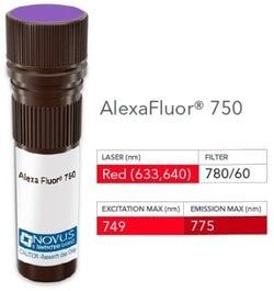
NB005744

Cytokeratin, LMW Antibody (AE-1), Alexa Fluor™ 350, Novus Biologicals™

Manufacturer: Novus Biologicals

Select a Size

Pack Size SKU Availability Price
Each of 1 NB005744-Each-of-1 In Stock ₹ 55,271.76

NB005744 - Each of 1

₹ 55,271.76

In Stock

Quantity

1

Base Price: ₹ 55,271.76

GST (18%): ₹ 9,948.917

Total Price: ₹ 65,220.677

Antigen

Cytokeratin, LMW

Classification

Monoclonal

Conjugate

Alexa Fluor 350

Formulation

50mM Sodium Borate with 0.05% Sodium Azide

Gene Symbols

KRT77

Immunogen

Human epidermal keratin (Uniprot: Q7Z794)

Quantity

0.1 mL

Primary or Secondary

Primary

Target Species

Human, Mouse, Rat, Bovine, Canine, Chicken, Primate, Rabbit

Isotype

IgG1 κ

Applications

Western Blot, Flow Cytometry, ELISA, Immunohistochemistry, Immunocytochemistry, Immunofluorescence, Immunohistochemistry (Paraffin)

Clone

AE-1

Dilution

Western Blot, Flow Cytometry, ELISA, Immunohistochemistry, Immunocytochemistry/Immunofluorescence, Immunohistochemistry-Paraffin, Immunohistochemistry-Frozen

Gene Alias

Cytokeratin LMW, cytokeratin-1B, keratin 77, keratin, type II cytoskeletal 1b, KRT77, type-II keratin Kb39

Host Species

Mouse

Purification Method

Protein A or G purified

Regulatory Status

RUO

Test Specificity

This monoclonal antibody recognizes the 56.5kDa (CK10); 50kDa (CK14); 50kDa (CK15); 48kDa (CK16); 40kDa (CK19) keratins of the acidic (Type I or LMW) subfamily. Twenty human keratins are resolved with two-dimensional gel electrophoresis into acidic (pI, 48, 46, 45, and 40kDa. monoclonal antibody AE3 recognizes the 65-67, 64, 59, 58, 56, and 52kDa keratins of basic subfamily. Many studies have shown the usefulness of keratins as markers in cancer research and tumor diagnosis. AE1/AE3 is a broad spectrum anti pan-keratin antibody cocktail, which differentiates epithelial tumors from non-epithelial tumors e.g. squamous vs. adenocarcinoma of the lung, liver carcinoma, breast cancer, and esophageal cancer.

Content And Storage

Store at 4°C in the dark.

Related Products

Img

Novus Biologicals

NB005748

--

Img

Novus Biologicals

NB005729

--

Img

Novus Biologicals

NB005732

--

Img

Novus Biologicals

NB005730

--

Img

Novus Biologicals

NB005733

--

Img

Novus Biologicals

NB005734

--

Img

Novus Biologicals

NB005769

--

Img

Novus Biologicals

NB005683

--

Description

  • Cytokeratin, LMW Monoclonal specifically detects Cytokeratin, LMW in Human, Mouse, Rat, Bovine, Canine, Chicken, Monkey, Rabbit, Reptile samples
  • It is validated for Western Blot, Flow Cytometry, Immunohistochemistry, Immunocytochemistry/Immunofluorescence, Immunohistochemistry-Paraffin.

Compare Similar Items

Show Difference

Img

Novus Biologicals

NB005744

--


Antigen:
Cytokeratin, LMW

Classification:
Monoclonal

Conjugate:
Alexa Fluor 350

Formulation:
50mM Sodium Borate with 0.05% Sodium Azide

Gene Symbols:
KRT77

Immunogen:
Human epidermal keratin (Uniprot: Q7Z794)

Quantity:
0.1 mL

Primary or Secondary:
Primary

Target Species:
Human, Mouse, Rat, Bovine, Canine, Chicken, Primate, Rabbit

Isotype:
IgG1 κ

Applications:
Western Blot, Flow Cytometry, ELISA, Immunohistochemistry, Immunocytochemistry, Immunofluorescence, Immunohistochemistry (Paraffin)

Clone:
AE-1

Dilution:
Western Blot, Flow Cytometry, ELISA, Immunohistochemistry, Immunocytochemistry/Immunofluorescence, Immunohistochemistry-Paraffin, Immunohistochemistry-Frozen

Gene Alias:
Cytokeratin LMW, cytokeratin-1B, keratin 77, keratin, type II cytoskeletal 1b, KRT77, type-II keratin Kb39

Host Species:
Mouse

Purification Method:
Protein A or G purified

Regulatory Status:
RUO

Test Specificity:
This monoclonal antibody recognizes the 56.5kDa (CK10); 50kDa (CK14); 50kDa (CK15); 48kDa (CK16); 40kDa (CK19) keratins of the acidic (Type I or LMW) subfamily. Twenty human keratins are resolved with two-dimensional gel electrophoresis into acidic (pI, 48, 46, 45, and 40kDa. monoclonal antibody AE3 recognizes the 65-67, 64, 59, 58, 56, and 52kDa keratins of basic subfamily. Many studies have shown the usefulness of keratins as markers in cancer research and tumor diagnosis. AE1/AE3 is a broad spectrum anti pan-keratin antibody cocktail, which differentiates epithelial tumors from non-epithelial tumors e.g. squamous vs. adenocarcinoma of the lung, liver carcinoma, breast cancer, and esophageal cancer.

Content And Storage:
Store at 4°C in the dark.

Img

Novus Biologicals

NB005748

--


Antigen:
Cytokeratin, LMW

Classification:
Monoclonal

Conjugate:
DyLight 594

Formulation:
50mM Sodium Borate with 0.05% Sodium Azide

Gene Symbols:
KRT77

Immunogen:
Human epidermal keratin (Uniprot: Q7Z794)

Quantity:
0.1 mL

Primary or Secondary:
Primary

Target Species:
Human, Mouse, Rat, Bovine, Canine, Chicken, Primate, Rabbit

Isotype:
IgG1 κ

Applications:
Western Blot, Flow Cytometry, ELISA, Immunohistochemistry, Immunocytochemistry, Immunofluorescence, Immunohistochemistry (Paraffin)

Clone:
AE-1

Dilution:
Western Blot, Flow Cytometry, ELISA, Immunohistochemistry, Immunocytochemistry/Immunofluorescence, Immunohistochemistry-Paraffin, Immunohistochemistry-Frozen

Gene Alias:
Cytokeratin LMW, cytokeratin-1B, keratin 77, keratin, type II cytoskeletal 1b, KRT77, type-II keratin Kb39

Host Species:
Mouse

Purification Method:
Protein A or G purified

Regulatory Status:
RUO

Test Specificity:
This monoclonal antibody recognizes the 56.5kDa (CK10); 50kDa (CK14); 50kDa (CK15); 48kDa (CK16); 40kDa (CK19) keratins of the acidic (Type I or LMW) subfamily. Twenty human keratins are resolved with two-dimensional gel electrophoresis into acidic (pI, 48, 46, 45, and 40kDa. monoclonal antibody AE3 recognizes the 65-67, 64, 59, 58, 56, and 52kDa keratins of basic subfamily. Many studies have shown the usefulness of keratins as markers in cancer research and tumor diagnosis. AE1/AE3 is a broad spectrum anti pan-keratin antibody cocktail, which differentiates epithelial tumors from non-epithelial tumors e.g. squamous vs. adenocarcinoma of the lung, liver carcinoma, breast cancer, and esophageal cancer.

Content And Storage:
Store at 4°C in the dark.

Img

Novus Biologicals

NB005750

--


Antigen:
TAG-72

Classification:
Monoclonal

Conjugate:
DyLight 594

Formulation:
50mM Sodium Borate with 0.05% Sodium Azide

Gene Symbols:
__

Immunogen:
A membrane enriched fraction of a human breast carcinoma liver metastasis was used as the immunogen for the TAG-72 (B72.3) antibody.

Quantity:
0.1 mL

Primary or Secondary:
Primary

Target Species:
Human, Rat, Bovine, Canine, Hamster

Isotype:
IgG1 κ

Applications:
Western Blot, Flow Cytometry, ELISA, Immunohistochemistry, Immunocytochemistry, Immunofluorescence, Immunohistochemistry (Paraffin)

Clone:
B72.3

Dilution:
Western Blot, Flow Cytometry, ELISA, Immunohistochemistry, Immunocytochemistry/Immunofluorescence, Immunohistochemistry-Paraffin, Immunohistochemistry-Frozen

Gene Alias:
CA 72-4, Protein TAG-72, tag 72, tag-72 Protein TAG-72

Host Species:
Mouse

Purification Method:
Protein A or G purified

Regulatory Status:
RUO

Test Specificity:
Recognizes an oncofetal antigen of 220kDa, identified as a tumor-associated glycoprotein (TAG-72) with properties of a mucin. This monoclonal antibody defines the mucin-carried sialylated-Tn epitope. TAG-72 is usually expressed by adenocarcinomas, but is negative in mesotheliomas. Studies have reported that this antibody has 80% sensitivity and 93% specificity for pulmonary adenocarcinoma. Therefore, TAG-72 is a useful marker to distinguish between mesothelioma and adenocarcinoma. However, false positive reactions can occur so results must be interpreted with the utmost caution. This antibody may be useful in the differentiation of non-small cell carcinomas from small cell carcinomas of the lung. The combined use of anti-TAG-72 and anti-GCDFP-15 is valuable in the diagnosis of apocrine carcinoma.

Content And Storage:
Store at 4°C in the dark.

Img

Novus Biologicals

NB005751

--


Antigen:
TAG-72

Classification:
Monoclonal

Conjugate:
FITC

Formulation:
PBS with 0.05% Sodium Azide

Gene Symbols:
__

Immunogen:
A membrane enriched fraction of a human breast carcinoma liver metastasis was used as the immunogen for the TAG-72 (B72.3) antibody.

Quantity:
0.1 mL

Primary or Secondary:
Primary

Target Species:
Human, Rat, Bovine, Canine, Hamster

Isotype:
IgG1 κ

Applications:
Flow Cytometry, ELISA, Immunohistochemistry, Immunocytochemistry, Immunofluorescence, Immunohistochemistry (Paraffin)

Clone:
B72.3

Dilution:
Flow Cytometry, ELISA, Immunohistochemistry, Immunocytochemistry/Immunofluorescence, Immunohistochemistry-Paraffin, Immunohistochemistry-Frozen

Gene Alias:
CA 72-4, Protein TAG-72, tag 72, tag-72 Protein TAG-72

Host Species:
Mouse

Purification Method:
Protein A or G purified

Regulatory Status:
RUO

Test Specificity:
Recognizes an oncofetal antigen of 220kDa, identified as a tumor-associated glycoprotein (TAG-72) with properties of a mucin. This monoclonal antibody defines the mucin-carried sialylated-Tn epitope. TAG-72 is usually expressed by adenocarcinomas, but is negative in mesotheliomas. Studies have reported that this antibody has 80% sensitivity and 93% specificity for pulmonary adenocarcinoma. Therefore, TAG-72 is a useful marker to distinguish between mesothelioma and adenocarcinoma. However, false positive reactions can occur so results must be interpreted with the utmost caution. This antibody may be useful in the differentiation of non-small cell carcinomas from small cell carcinomas of the lung. The combined use of anti-TAG-72 and anti-GCDFP-15 is valuable in the diagnosis of apocrine carcinoma.

Content And Storage:
Store at 4°C in the dark.