NB005757

Cytokeratin 17 Antibody (E3 (same as Ks17.E3)), Alexa Fluor™ 750, Novus Biologicals™

Manufacturer: Novus Biologicals

Select a Size

Pack Size SKU Availability Price
Each of 1 NB005757-Each-of-1 In Stock ₹ 55,271.76

NB005757 - Each of 1

₹ 55,271.76

In Stock

Quantity

1

Base Price: ₹ 55,271.76

GST (18%): ₹ 9,948.917

Total Price: ₹ 65,220.677

Antigen

Cytokeratin 17

Classification

Monoclonal

Conjugate

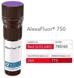
Alexa Fluor 750

Formulation

50mM Sodium Borate with 0.05% Sodium Azide

Gene Symbols

KRT17

Immunogen

Cytoskeletal fraction of rat colon epithelium (Uniprot: Q04695)

Quantity

0.1 mL

Research Discipline

Cancer

Test Specificity

Cytokeratin 17 (CK17) is normally expressed in the basal cells of complex epithelia but not in stratified or simple epithelia. Antibody to CK17 is an excellent tool to distinguish myoepithelial cells from luminal epithelium of various glands such as mammary, sweat and salivary. CK17 is expressed in epithelial cells of various origins, such as bronchial epithelial cells and skin appendages. It may be considered as 'epithelial stem cell' marker because CK17 Ab marks basal cell differentiation. CK17 is expressed in SCLC much higher than in LADC. Eighty-five percent of the triple negative breast carcinomas immunoreact with basal cytokeratins including anti-CK17. Also important is that cases of triple negative breast carcinoma with expression of CK17 show an aggressive clinical course. The histologic differentiation of ampullary cancer, intestinal vs. pancreatobiliary, is very important for treatment. Usually anti-CK17 and anti-MUC1 immunoreactivity represents pancreatobiliary subtype whereas anti-MUC2 and anti-CDX-2 positivity defines intestinal subtype.

Content And Storage

Store at 4°C in the dark.

Applications

Western Blot, ELISA, Immunocytochemistry, Immunofluorescence, Immunohistochemistry (Paraffin)

Clone

E3 (same as Ks17.E3)

Dilution

Western Blot, ELISA, Immunocytochemistry/Immunofluorescence, Immunohistochemistry-Paraffin

Gene Alias

39.1, CK-17, cytokeratin-17, K17, keratin 17, keratin, type I cytoskeletal 17, keratin-17, PC, PC2, PCHC1

Host Species

Mouse

Purification Method

Protein A or G purified

Regulatory Status

RUO

Primary or Secondary

Primary

Target Species

Human, Rat, Porcine, Bovine, Goat

Isotype

IgG2b κ

Related Products

Img

Novus Biologicals

NB006261

--

Img

Novus Biologicals

NB005813

--

Img

Novus Biologicals

NB005812

--

Img

Novus Biologicals

NB006138

--

Img

Novus Biologicals

NB005616

--

Img

Novus Biologicals

NB005743

--

Img

Novus Biologicals

NB005808

--

Img

Novus Biologicals

NB007162

--

Description

  • Cytokeratin 17 Monoclonal specifically detects Cytokeratin 17 in Human, Rat, Porcine, Bovine, Goat samples
  • It is validated for Western Blot, Flow Cytometry, Immunocytochemistry/Immunofluorescence, Immunohistochemistry-Paraffin, Immunohistochemistry-Frozen.

Compare Similar Items

Show Difference

Img

Novus Biologicals

NB005757

--


Antigen:
Cytokeratin 17

Classification:
Monoclonal

Conjugate:
Alexa Fluor 750

Formulation:
50mM Sodium Borate with 0.05% Sodium Azide

Gene Symbols:
KRT17

Immunogen:
Cytoskeletal fraction of rat colon epithelium (Uniprot: Q04695)

Quantity:
0.1 mL

Research Discipline:
Cancer

Test Specificity:
Cytokeratin 17 (CK17) is normally expressed in the basal cells of complex epithelia but not in stratified or simple epithelia. Antibody to CK17 is an excellent tool to distinguish myoepithelial cells from luminal epithelium of various glands such as mammary, sweat and salivary. CK17 is expressed in epithelial cells of various origins, such as bronchial epithelial cells and skin appendages. It may be considered as 'epithelial stem cell' marker because CK17 Ab marks basal cell differentiation. CK17 is expressed in SCLC much higher than in LADC. Eighty-five percent of the triple negative breast carcinomas immunoreact with basal cytokeratins including anti-CK17. Also important is that cases of triple negative breast carcinoma with expression of CK17 show an aggressive clinical course. The histologic differentiation of ampullary cancer, intestinal vs. pancreatobiliary, is very important for treatment. Usually anti-CK17 and anti-MUC1 immunoreactivity represents pancreatobiliary subtype whereas anti-MUC2 and anti-CDX-2 positivity defines intestinal subtype.

Content And Storage:
Store at 4°C in the dark.

Applications:
Western Blot, ELISA, Immunocytochemistry, Immunofluorescence, Immunohistochemistry (Paraffin)

Clone:
E3 (same as Ks17.E3)

Dilution:
Western Blot, ELISA, Immunocytochemistry/Immunofluorescence, Immunohistochemistry-Paraffin

Gene Alias:
39.1, CK-17, cytokeratin-17, K17, keratin 17, keratin, type I cytoskeletal 17, keratin-17, PC, PC2, PCHC1

Host Species:
Mouse

Purification Method:
Protein A or G purified

Regulatory Status:
RUO

Primary or Secondary:
Primary

Target Species:
Human, Rat, Porcine, Bovine, Goat

Isotype:
IgG2b κ

Img

Novus Biologicals

NB005761

--


Antigen:
Bcl-10

Classification:
Monoclonal

Conjugate:
FITC

Formulation:
PBS with 0.05% Sodium Azide

Gene Symbols:
BCL10

Immunogen:
Human Bcl-10 recombinant protein (epitope aa122-168) (Uniprot: O95999)

Quantity:
0.1 mL

Research Discipline:
Apoptosis, Cancer

Test Specificity:
BCL10, with an N-terminal caspase recruitment domain (CARD), is found in a number of apoptotic regulatory molecules. It was identified through its direct involvement in t(1;14) of mucosa-associated lymphoid tissue (MALT) lymphoma. Expression of BCL10 was shown to induce NFKB activation in a NIK-dependent pathway. This MAb labels subpopulations of normal B and T cells and is a useful tool for the sub-classification of lymphomas. In MALT lymphomas with the t(1;14) translocation, while 55% of MALT lymphomas lacking this translocation exhibited the same labeling pattern, although at a much lower level.

Content And Storage:
Store at 4°C in the dark.

Applications:
ELISA

Clone:
BL10/411

Dilution:
ELISA

Gene Alias:
B-cell CLL/lymphoma 10CIPERCLAPB-cell lymphoma/leukemia 10, Bcl-10, CARD-containing apoptotic signaling protein, CARD-containing molecule enhancing NF-kappa-B, CARD-containing proapoptotic protein, CARD-like apoptotic protein, CARMEN, caspase-recruiting domain-containing protein, cCARMEN, c-E10bcl-10, CED-3/ICH-1 prodomain homologous E10-like regulator, Cellular homolog of vCARMEN, cellular-E10, hCLAP, Mammalian CARD-containing adapter molecule E10, mE10CARD containing molecule enhancing NF-kB

Host Species:
Mouse

Purification Method:
Protein A or G purified

Regulatory Status:
RUO

Primary or Secondary:
Primary

Target Species:
Human

Isotype:
IgG1 κ

Img

Novus Biologicals

NB005764

--


Antigen:
CD19

Classification:
Monoclonal

Conjugate:
DyLight 594

Formulation:
50mM Sodium Borate with 0.05% Sodium Azide

Gene Symbols:
CD19

Immunogen:
Recombinant full-length human CD19 protein (Uniprot: P15391)

Quantity:
0.1 mL

Research Discipline:
Adaptive Immunity, B Cell Development and Differentiation Markers, Cytokine Research, Immunology, Innate Immunity, Mesenchymal Stem Cell Markers, Myeloid derived Suppressor Cell, Signal Transduction, Stem Cell Lines, Stem Cell Markers

Test Specificity:
__

Content And Storage:
Store at 4°C in the dark.

Applications:
Immunohistochemistry, Immunohistochemistry (Paraffin)

Clone:
C19/366

Dilution:
Immunohistochemistry, Immunohistochemistry-Paraffin

Gene Alias:
B4, B-lymphocyte antigen CD19, B-lymphocyte surface antigen B4, CD19 antigen, CD19 molecule, CVID3, Differentiation antigen CD19, MGC12802, T-cell surface antigen Leu-12

Host Species:
Mouse

Purification Method:
Protein A or G purified

Regulatory Status:
RUO

Primary or Secondary:
Primary

Target Species:
Human

Isotype:
IgG1 κ

Img

Novus Biologicals

NB005765

--


Antigen:
CD30/TNFRSF8

Classification:
Monoclonal

Conjugate:
DyLight 594

Formulation:
50mM Sodium Borate with 0.05% Sodium Azide

Gene Symbols:
TNFRSF8

Immunogen:
Human CD30/TNFRSF8 recombinant protein (Uniprot: P28908)

Quantity:
0.1 mL

Research Discipline:
Apoptosis, B Cell Development and Differentiation Markers, Cell Cycle and Replication, Immunology, Stem Cell Markers

Test Specificity:
Recognizes a single chain glycoprotein of 105/120kDa, identified as CD30/Ki-1. CD30 is synthesized as a 90kDa precursor, which is processed in the Golgi complex into a membrane-bound phosphorylated mature 105/120kDa glycoprotein. In Hodgkins disease, CD30/Ki-1 antigen is expressed by mononuclear-Hodgkin and multinucleated Reed-Sternberg cells. It is also expressed by the tumor cells of a majority of anaplastic large cell lymphomas as well as by a varying proportion of activated T and B cells. This monoclonal antibody distinguishes large cell lymphomas derived from activated lymphoid cells from histiocytic malignancies and lymphomas derived from resting and precursor lymphoid cells or from anaplastic carcinomas. About one third of the Ki-1 positive lymphomas lack the leukocyte common antigen (CD45).

Content And Storage:
Store at 4°C in the dark.

Applications:
Flow Cytometry, ELISA, Immunohistochemistry, Immunocytochemistry, Immunofluorescence, Immunohistochemistry (Paraffin)

Clone:
CD30/412

Dilution:
Flow Cytometry, ELISA, Immunohistochemistry, Immunocytochemistry/Immunofluorescence, Immunohistochemistry-Paraffin, Immunohistochemistry-Frozen

Gene Alias:
CD30, CD30 antigen, CD30KI-1, CD30L receptor, cytokine receptor CD30, D1S166EKi-1, Ki-1 antigen, Lymphocyte activation antigen CD30, tumor necrosis factor receptor superfamily member 8, tumor necrosis factor receptor superfamily, member 8

Host Species:
Mouse

Purification Method:
Protein A or G purified

Regulatory Status:
RUO

Primary or Secondary:
Primary

Target Species:
Human

Isotype:
IgG1 κ