NB006238

Myogenin Antibody (F5D), FITC, Novus Biologicals™

Manufacturer: Novus Biologicals

Select a Size

Pack Size SKU Availability Price
Each of 1 NB006238-Each-of-1 In Stock ₹ 55,271.76

NB006238 - Each of 1

₹ 55,271.76

In Stock

Quantity

1

Base Price: ₹ 55,271.76

GST (18%): ₹ 9,948.917

Total Price: ₹ 65,220.677

Antigen

Myogenin

Classification

Monoclonal

Conjugate

FITC

Formulation

PBS with 0.05% Sodium Azide

Gene Symbols

MYOG

Immunogen

Rat Myogenin peptide (aa 73-94) followed by rat Myogenin recombinant fragment (aa30-224) (Epitope aa138-158) (Uniprot: P15173)

Quantity

0.1 mL

Research Discipline

Cancer, Stem Cell Markers, Transcription Factors and Regulators

Test Specificity

Myogenin is a member of the MyoD family of myogenic basic helix-loop-helix (bHLH) transcription factors that also includes MyoD, Myf-5, and MRF4 (also known as herculinor Myf-6). MyoD family members are expressed exclusively in skeletal muscle and play a key role in activating myogenesis by binding to enhancer sequences of muscle-specific genes. The regulatory domain of MyoD is approximately 70 amino acids in length and includes both a basic DNA binding motif and a bHLH dimerization motif. MyoD family members share about 80% amino acid homology in their bHLH motifs.Anti-myogenin labels the nuclei of myoblasts in developing muscle tissue, and is expressed in tumor cell nuclei of rhabdomyosarcoma and some leiomyosarcomas. Positive nuclear staining may occur in Wilms' tumor.

Content And Storage

Store at 4°C in the dark.

Applications

Flow Cytometry, ELISA, Immunohistochemistry, Immunocytochemistry, Immunofluorescence, Immunohistochemistry (Paraffin)

Clone

F5D

Dilution

Flow Cytometry, ELISA, Immunohistochemistry, Immunocytochemistry/Immunofluorescence, Immunohistochemistry-Paraffin, Immunohistochemistry-Frozen

Gene Alias

BHLHC3, bHLHc3Myogenic factor-4; myogenin, Class C basic helix-loop-helix protein 3, myf-4, MYF4myogenin, Myogenic factor 4, MYOGENIN, myogenin (myogenic factor 4)

Host Species

Mouse

Purification Method

Protein A or G purified

Regulatory Status

RUO

Primary or Secondary

Primary

Target Species

Human, Mouse, Rat, Porcine, Feline

Isotype

IgG1 κ

Related Products

Img

Novus Biologicals

NB006329

--

Img

Novus Biologicals

NP247917A32

--

Img

Novus Biologicals

NB006796

--

Img

Novus Biologicals

N247901J646

--

Img

Novus Biologicals

NP247901A70

--

Img

Novus Biologicals

NB10056511K

--

Img

Novus Biologicals

NB006336

--

Img

Novus Biologicals

NP24788A32

--

Description

  • Myogenin Monoclonal specifically detects Myogenin in Human, Mouse, Rat, Porcine, Canine, Feline samples
  • It is validated for ELISA, Immunohistochemistry, Immunohistochemistry-Paraffin.

Compare Similar Items

Show Difference

Img

Novus Biologicals

NB006238

--


Antigen:
Myogenin

Classification:
Monoclonal

Conjugate:
FITC

Formulation:
PBS with 0.05% Sodium Azide

Gene Symbols:
MYOG

Immunogen:
Rat Myogenin peptide (aa 73-94) followed by rat Myogenin recombinant fragment (aa30-224) (Epitope aa138-158) (Uniprot: P15173)

Quantity:
0.1 mL

Research Discipline:
Cancer, Stem Cell Markers, Transcription Factors and Regulators

Test Specificity:
Myogenin is a member of the MyoD family of myogenic basic helix-loop-helix (bHLH) transcription factors that also includes MyoD, Myf-5, and MRF4 (also known as herculinor Myf-6). MyoD family members are expressed exclusively in skeletal muscle and play a key role in activating myogenesis by binding to enhancer sequences of muscle-specific genes. The regulatory domain of MyoD is approximately 70 amino acids in length and includes both a basic DNA binding motif and a bHLH dimerization motif. MyoD family members share about 80% amino acid homology in their bHLH motifs.Anti-myogenin labels the nuclei of myoblasts in developing muscle tissue, and is expressed in tumor cell nuclei of rhabdomyosarcoma and some leiomyosarcomas. Positive nuclear staining may occur in Wilms' tumor.

Content And Storage:
Store at 4°C in the dark.

Applications:
Flow Cytometry, ELISA, Immunohistochemistry, Immunocytochemistry, Immunofluorescence, Immunohistochemistry (Paraffin)

Clone:
F5D

Dilution:
Flow Cytometry, ELISA, Immunohistochemistry, Immunocytochemistry/Immunofluorescence, Immunohistochemistry-Paraffin, Immunohistochemistry-Frozen

Gene Alias:
BHLHC3, bHLHc3Myogenic factor-4; myogenin, Class C basic helix-loop-helix protein 3, myf-4, MYF4myogenin, Myogenic factor 4, MYOGENIN, myogenin (myogenic factor 4)

Host Species:
Mouse

Purification Method:
Protein A or G purified

Regulatory Status:
RUO

Primary or Secondary:
Primary

Target Species:
Human, Mouse, Rat, Porcine, Feline

Isotype:
IgG1 κ

Img

Novus Biologicals

NB006244

--


Antigen:
FoxP3

Classification:
Monoclonal

Conjugate:
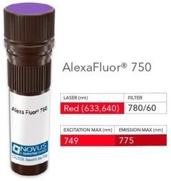
Alexa Fluor 750

Formulation:
50mM Sodium Borate with 0.05% Sodium Azide

Gene Symbols:
FOXP3

Immunogen:
Recombinant human full-length FoxP3 protein (Uniprot: Q9ZS1)

Quantity:
0.1 mL

Research Discipline:
Adaptive Immunity, Cell Biology, Chromatin Research, Immunology, Regulatory Immunology, Transcription Factors and Regulators

Test Specificity:
Recognizes a protein of 47-55kDa, which is identified as FOXP3. Its precise epitope is not known, but it has been mapped to the N-terminal portion of the protein. The FOX family of transcription factors is a large group of proteins that share a common DNA binding domain termed a winged-helix or forkhead domain. During early development, FOXP1 and FOXP2 are expressed abundantly in the lung, with lower levels of expression in neural, intestinal and cardiovascular tissues, where they act as transcription repressors. FOXP1 is widely expressed in adult tissues, while neoplastic cells often exhibit a dramatic change in expression level or localization of FOXP1. Mutations in FOXP3 gene cause IPEX, a fatal, X-linked inherited disorder characterized by immune dysregulation. The FOXP3 protein is essential for normal immune homeostasis. Specifically, FOXP3 represses transcription through a DNA binding forkhead domain, thereby regulating T cell activation.

Content And Storage:
Store at 4°C in the dark.

Applications:
Flow Cytometry, ELISA, Immunohistochemistry, Immunocytochemistry, Immunofluorescence, Immunohistochemistry (Paraffin), Immunohistochemistry (Frozen)

Clone:
FXP3/197

Dilution:
Flow Cytometry, ELISA, Immunohistochemistry, Immunocytochemistry/Immunofluorescence, Immunohistochemistry-Paraffin, Immunohistochemistry-Frozen

Gene Alias:
AIID, AIIDMGC141961, DIETER, forkhead box P3, Forkhead Box Protein P3, FOXP3delta7, immune dysregulation, polyendocrinopathy, enteropathy, X-linked, Immunodeficiency, Polyendocrinopathy, Enteropathy, X-Linked, IPEX, JM2, MGC141961, MGC141963, PIDX, PIDXMGC141963, Scurfin, XPID, XPIDpolyendocrinopathy, enteropathy, X-linked

Host Species:
Mouse

Purification Method:
Protein A or G purified

Regulatory Status:
RUO

Primary or Secondary:
Primary

Target Species:
Human, Mouse, Primate

Isotype:
IgG1 κ

Img

Novus Biologicals

NB006245

--


Antigen:
FoxP3

Classification:
Monoclonal

Conjugate:
FITC

Formulation:
PBS with 0.05% Sodium Azide

Gene Symbols:
FOXP3

Immunogen:
Recombinant human full-length FoxP3 protein (Uniprot: Q9ZS1)

Quantity:
0.1 mL

Research Discipline:
Adaptive Immunity, Cell Biology, Chromatin Research, Immunology, Regulatory Immunology, Transcription Factors and Regulators

Test Specificity:
Recognizes a protein of 47-55kDa, which is identified as FOXP3. Its precise epitope is not known, but it has been mapped to the N-terminal portion of the protein. The FOX family of transcription factors is a large group of proteins that share a common DNA binding domain termed a winged-helix or forkhead domain. During early development, FOXP1 and FOXP2 are expressed abundantly in the lung, with lower levels of expression in neural, intestinal and cardiovascular tissues, where they act as transcription repressors. FOXP1 is widely expressed in adult tissues, while neoplastic cells often exhibit a dramatic change in expression level or localization of FOXP1. Mutations in FOXP3 gene cause IPEX, a fatal, X-linked inherited disorder characterized by immune dysregulation. The FOXP3 protein is essential for normal immune homeostasis. Specifically, FOXP3 represses transcription through a DNA binding forkhead domain, thereby regulating T cell activation.

Content And Storage:
Store at 4°C in the dark.

Applications:
Flow Cytometry, ELISA, Immunohistochemistry, Immunocytochemistry, Immunofluorescence, Immunohistochemistry (Paraffin), Immunohistochemistry (Frozen)

Clone:
FXP3/197

Dilution:
Flow Cytometry, ELISA, Immunohistochemistry, Immunocytochemistry/Immunofluorescence, Immunohistochemistry-Paraffin, Immunohistochemistry-Frozen

Gene Alias:
AIID, AIIDMGC141961, DIETER, forkhead box P3, Forkhead Box Protein P3, FOXP3delta7, immune dysregulation, polyendocrinopathy, enteropathy, X-linked, Immunodeficiency, Polyendocrinopathy, Enteropathy, X-Linked, IPEX, JM2, MGC141961, MGC141963, PIDX, PIDXMGC141963, Scurfin, XPID, XPIDpolyendocrinopathy, enteropathy, X-linked

Host Species:
Mouse

Purification Method:
Protein A or G purified

Regulatory Status:
RUO

Primary or Secondary:
Primary

Target Species:
Human, Mouse, Primate

Isotype:
IgG1 κ

Img

Novus Biologicals

NB006248

--


Antigen:
HSP27

Classification:
Monoclonal

Conjugate:
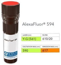
Alexa Fluor 594

Formulation:
50mM Sodium Borate with 0.05% Sodium Azide

Gene Symbols:
HSPB1

Immunogen:
Partially purified hsp27 (earlier called 24K) protein from breast cancer MCF-7 cells.

Quantity:
0.1 mL

Research Discipline:
Cancer, Cell Biology, Cell Cycle and Replication, Golgi Apparatus Markers, Membrane Trafficking and Chaperones, Neuroscience, Phospho Specific

Test Specificity:
monoclonal antibody G3.1 reacts specifically with heat shock protein HSP27 in human and monkey tissues and cell lines such as MCF-7. HSP27, also referred to as the Estrogen-Regulated 24K protein and HSP28, is one of several small heat shock proteins produced by all organisms studied. HSP27 synthesis is induced by elevated temperature, as well as by estrogen in hormone responsive cells. Interestingly, human HSP27 also shares greater than 50% homology with low molecular weight Drosophila HSPs and mammalian alpha-crystalline lens protein. Because of the estrogen responsive nature of HSP27, this protein has been studied extensively in human estrogen responsive tissues such as cervix, endometrium and breast tissue. Therefore, HSP27 may be useful in classifying various hormone sensitive tumors.

Content And Storage:
Store at 4°C in the dark.

Applications:
Western Blot, Flow Cytometry, ELISA, Immunohistochemistry, Immunocytochemistry, Immunofluorescence, Immunohistochemistry (Paraffin)

Clone:
G3.1

Dilution:
Western Blot, Flow Cytometry, ELISA, Immunohistochemistry, Immunocytochemistry/Immunofluorescence, Immunohistochemistry-Paraffin, Immunohistochemistry-Frozen

Gene Alias:
28 kDa heat shock protein, DKFZp586P1322, Estrogen-regulated 24 kDa protein, Heat shock 27 kDa protein, heat shock 27kD protein 1, heat shock 27kDa protein 1, heat shock protein beta-1, HMN2B, HS.76067, Hsp25, HSP27HSP 27, HSP28CMT2F, HspB1, SRP27, Stress-responsive protein 27

Host Species:
Mouse

Purification Method:
Protein A or G purified

Regulatory Status:
RUO

Primary or Secondary:
Primary

Target Species:
Human, Mouse, Rat, Chicken, Primate, Sheep

Isotype:
IgG1 κ