ErbB2/Her2 Antibody (HRB2/282), FITC, Novus Biologicals™
Manufacturer: Novus Biologicals
Select a Size
| Pack Size | SKU | Availability | Price |
|---|---|---|---|
| Each of 1 | NB006280-Each-of-1 | In Stock | ₹ 59,674.50 |
NB006280 - Each of 1
In Stock
Quantity
1
Base Price: ₹ 59,674.50
GST (18%): ₹ 10,741.41
Total Price: ₹ 70,415.91
Antigen
ErbB2/Her2
Classification
Monoclonal
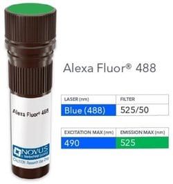
Conjugate
FITC
Formulation
PBS with 0.05% Sodium Azide
Gene Symbols
ERBB2
Immunogen
Recombinant extracellular domain of human ErbB2/Her2 protein (Uniprot: P04626)
Quantity
0.1 mL
Research Discipline
Breast Cancer, Cancer, Cellular Markers, Core ESC Like Genes, Oncogenes, Phospho Specific, Protein Kinase, Stem Cell Markers, Tumor Suppressors
Test Specificity
Recognizes a protein of 185kDa, which is identified as c-erbB-2/HER-2/neu. Its epitope is localized in the extracellular domain. C-erbB-2/HER-2 is a member of the EGFR family. This monoclonal antibody is specific and shows minimal cross-reaction with other members of the EGFR-family. Receptors of this family are located on the plasma membrane and consist of an extracellular ligand-binding domain that is connected to a large intracellular domain by a single transmembrane sequence. c-erbB-2/HER-2 protein is over-expressed in a variety of carcinomas especially those of breast and ovary.
Content And Storage
Store at 4°C in the dark.
Applications
Flow Cytometry, ELISA, Immunohistochemistry, Immunocytochemistry, Immunofluorescence, Immunohistochemistry (Frozen)
Clone
HRB2/282
Dilution
Flow Cytometry, ELISA, Immunohistochemistry, Immunocytochemistry/Immunofluorescence, Immunohistochemistry-Frozen
Gene Alias
CD340, CD340 antigen, c-erb B2/neu protein, EC 2.7.10, EGFR2, HER-2, HER2EC 2.7.10.1, herstatin, Metastatic lymph node gene 19 protein, MLN 19, MLN19, NEUHER-2/neu, neuroblastoma/glioblastoma derived oncogene homolog, NGLTKR1, p185erbB2, Proto-oncogene c-ErbB-2, Proto-oncogene Neu, receptor tyrosine-protein kinase erbB-2, Tyrosine kinase-type cell surface receptor HER2, v-erb-b2 avian erythroblastic leukemia viral oncogene homolog 2(neuro/glioblastoma derived oncogene homolog), v-erb-b2 erythroblastic leukemia viral oncogene homolog 2, neuro/glioblastomaderived oncogene homolog (avian)
Host Species
Mouse
Purification Method
Protein A or G purified
Regulatory Status
RUO
Primary or Secondary
Primary
Target Species
Human
Isotype
IgG1 κ
Related Products

Description
- ErbB2/Her2 Monoclonal specifically detects ErbB2/Her2 in Human samples
- It is validated for Flow Cytometry, ELISA, Immunocytochemistry/Immunofluorescence.
Compare Similar Items
Show Difference

Antigen: ErbB2/Her2
Classification: Monoclonal
Conjugate: FITC
Formulation: PBS with 0.05% Sodium Azide
Gene Symbols: ERBB2
Immunogen: Recombinant extracellular domain of human ErbB2/Her2 protein (Uniprot: P04626)
Quantity: 0.1 mL
Research Discipline: Breast Cancer, Cancer, Cellular Markers, Core ESC Like Genes, Oncogenes, Phospho Specific, Protein Kinase, Stem Cell Markers, Tumor Suppressors
Test Specificity: Recognizes a protein of 185kDa, which is identified as c-erbB-2/HER-2/neu. Its epitope is localized in the extracellular domain. C-erbB-2/HER-2 is a member of the EGFR family. This monoclonal antibody is specific and shows minimal cross-reaction with other members of the EGFR-family. Receptors of this family are located on the plasma membrane and consist of an extracellular ligand-binding domain that is connected to a large intracellular domain by a single transmembrane sequence. c-erbB-2/HER-2 protein is over-expressed in a variety of carcinomas especially those of breast and ovary.
Content And Storage: Store at 4°C in the dark.
Applications: Flow Cytometry, ELISA, Immunohistochemistry, Immunocytochemistry, Immunofluorescence, Immunohistochemistry (Frozen)
Clone: HRB2/282
Dilution: Flow Cytometry, ELISA, Immunohistochemistry, Immunocytochemistry/Immunofluorescence, Immunohistochemistry-Frozen
Gene Alias: CD340, CD340 antigen, c-erb B2/neu protein, EC 2.7.10, EGFR2, HER-2, HER2EC 2.7.10.1, herstatin, Metastatic lymph node gene 19 protein, MLN 19, MLN19, NEUHER-2/neu, neuroblastoma/glioblastoma derived oncogene homolog, NGLTKR1, p185erbB2, Proto-oncogene c-ErbB-2, Proto-oncogene Neu, receptor tyrosine-protein kinase erbB-2, Tyrosine kinase-type cell surface receptor HER2, v-erb-b2 avian erythroblastic leukemia viral oncogene homolog 2(neuro/glioblastoma derived oncogene homolog), v-erb-b2 erythroblastic leukemia viral oncogene homolog 2, neuro/glioblastomaderived oncogene homolog (avian)
Host Species: Mouse
Purification Method: Protein A or G purified
Regulatory Status: RUO
Primary or Secondary: Primary
Target Species: Human
Isotype: IgG1 κ
Antigen: Hepatocyte Specific Antigen
Classification: Monoclonal
Conjugate: FITC
Formulation: PBS with 0.05% Sodium Azide
Gene Symbols: __
Immunogen: SK-H1A9-2 human hepatocellular carcinoma cells
Quantity: 0.1 mL
Research Discipline: __
Test Specificity: monoclonal antibody HSA133 stains human liver canaliculi and a subset of hepatocellular carcinomas. In frozen sections, it stains liver canaliculi strongly and may be used as a marker of this hepatic substructure. Cell preparations of hepatocellular carcinoma biopsies and cell lines are found to bind this monoclonal antibody on the cell surface. HSA133 strongly stains liver canaliculi and hepatic carcinoma cells using frozen sections or paraformaldehyde fixed cell preparations.
Content And Storage: Store at 4°C in the dark.
Applications: Immunocytochemistry, Immunofluorescence
Clone: HSA133
Dilution: Immunocytochemistry/Immunofluorescence
Gene Alias: Hepatocyte, HSA
Host Species: Mouse
Purification Method: Protein A or G purified
Regulatory Status: RUO
Primary or Secondary: Primary
Target Species: Human
Isotype: IgG2b κ

Antigen: Biotin (Vitamin B7 or Vitamin H)
Classification: Monoclonal
Conjugate: DyLight 594
Formulation: 50mM Sodium Borate with 0.05% Sodium Azide
Gene Symbols: __
Immunogen: Biotinylated sheep immunoglobulin
Quantity: 0.1 mL
Research Discipline: __
Test Specificity: It recognizes both the free and protein-conjugated (either soluble or cell bound) form of biotin. This monoclonal antibody is highly specific to biotin and shows no cross-reaction with other structurally related compounds. It has a very high affinity for biotin and is excellent for use in various amplification techniques. In some applications, localization of biotinylated probes with avidin produces unacceptably high background staining. Anti-biotin antibody may be substituted to decrease this noise.
Content And Storage: Store at 4°C in the dark.
Applications: Western Blot, Flow Cytometry, ELISA, Immunohistochemistry, Immunocytochemistry, Immunofluorescence, Immunohistochemistry (Paraffin)
Clone: Hyb-8
Dilution: Western Blot, Flow Cytometry, ELISA, Immunohistochemistry, Immunocytochemistry/Immunofluorescence, Immunohistochemistry-Paraffin, Immunohistochemistry-Frozen
Gene Alias: 58-85-5, coenzyme R, d-biotin, Vitamin B7, vitamin H
Host Species: Mouse
Purification Method: Protein A or G purified
Regulatory Status: RUO
Primary or Secondary: Primary
Target Species: All species
Isotype: IgG1 κ