NB006449

MUC1 Antibody (SPM132), Alexa Fluor™ 750, Novus Biologicals™

Manufacturer: Novus Biologicals

Select a Size

Pack Size SKU Availability Price
Each of 1 NB006449-Each-of-1 In Stock ₹ 55,271.76

NB006449 - Each of 1

₹ 55,271.76

In Stock

Quantity

1

Base Price: ₹ 55,271.76

GST (18%): ₹ 9,948.917

Total Price: ₹ 65,220.677

Antigen

MUC1

Classification

Monoclonal

Conjugate

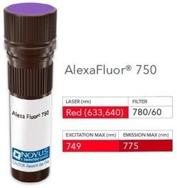
Alexa Fluor 750

Formulation

50mM Sodium Borate with 0.05% Sodium Azide

Gene Symbols

MUC1

Immunogen

Human milk fat globule membranes (Uniprot: P15941)

Quantity

0.1 mL

Research Discipline

Cancer, Cellular Markers, Extracellular Matrix, Inflammation, Signal Transduction

Test Specificity

In Western blotting, it recognizes proteins in MW range of 265-400kDa, identified as different glycoforms of EMA. This monoclonal antibody reacts with the DTRP epitope in the tandem repeats. The alpha subunit has cell adhesive properties. It can act both as an adhesion and an anti-adhesion protein. EMA may provide a protective layer on epithelial cells against bacterial and enzyme attack. The beta subunit contains a C-terminal domain, which is involved in cell signaling, through phosphorylations and protein-protein interactions. In immunohistochemical assays, it superbly stains routine formalin/paraffin carcinoma tissues. Antibody to EMA is useful as a pan-epithelial marker for detecting early metastatic loci of carcinoma in bone marrow or liver.

Content And Storage

Store at 4°C in the dark.

Applications

Western Blot, Flow Cytometry, ELISA, Immunohistochemistry, Immunocytochemistry, Immunofluorescence, Immunohistochemistry (Paraffin)

Clone

SPM132

Dilution

Western Blot, Flow Cytometry, ELISA, Immunohistochemistry, Immunocytochemistry/Immunofluorescence, Immunohistochemistry-Paraffin, Immunohistochemistry-Frozen

Gene Alias

Breast carcinoma-associated antigen DF3, Carcinoma-associated mucin, CD227, CD227 antigen, DF3 antigen, EMA, episialin, H23 antigen, H23AG, KL-6, MAM6, MUC-1, MUC1/ZD, mucin 1, cell surface associated, mucin 1, transmembrane, mucin-1, Peanut-reactive urinary mucin, PEMMUC-1/SEC, PEMT, Polymorphic epithelial mucin, PUMMUC-1/X, tumor associated epithelial mucin, Tumor-associated epithelial membrane antigen, Tumor-associated mucin

Host Species

Mouse

Purification Method

Protein A or G purified

Regulatory Status

RUO

Primary or Secondary

Primary

Target Species

Human

Isotype

IgG1 κ

Related Products

Img

Novus Biologicals

NB006446

--

Img

Novus Biologicals

NB006447

--

Img

Novus Biologicals

NB006702

--

Img

Novus Biologicals

NB006699

--

Img

Novus Biologicals

NB006701

--

Img

Novus Biologicals

NB006254

--

Img

Novus Biologicals

NB006703

--

Img

Novus Biologicals

NB006704

--

Description

  • MUC1 Monoclonal specifically detects MUC1 in Human samples
  • It is validated for Western Blot, Flow Cytometry, Immunohistochemistry, Immunocytochemistry/Immunofluorescence, Immunohistochemistry-Paraffin.

Compare Similar Items

Show Difference

Img

Novus Biologicals

NB006449

--


Antigen:
MUC1

Classification:
Monoclonal

Conjugate:
Alexa Fluor 750

Formulation:
50mM Sodium Borate with 0.05% Sodium Azide

Gene Symbols:
MUC1

Immunogen:
Human milk fat globule membranes (Uniprot: P15941)

Quantity:
0.1 mL

Research Discipline:
Cancer, Cellular Markers, Extracellular Matrix, Inflammation, Signal Transduction

Test Specificity:
In Western blotting, it recognizes proteins in MW range of 265-400kDa, identified as different glycoforms of EMA. This monoclonal antibody reacts with the DTRP epitope in the tandem repeats. The alpha subunit has cell adhesive properties. It can act both as an adhesion and an anti-adhesion protein. EMA may provide a protective layer on epithelial cells against bacterial and enzyme attack. The beta subunit contains a C-terminal domain, which is involved in cell signaling, through phosphorylations and protein-protein interactions. In immunohistochemical assays, it superbly stains routine formalin/paraffin carcinoma tissues. Antibody to EMA is useful as a pan-epithelial marker for detecting early metastatic loci of carcinoma in bone marrow or liver.

Content And Storage:
Store at 4°C in the dark.

Applications:
Western Blot, Flow Cytometry, ELISA, Immunohistochemistry, Immunocytochemistry, Immunofluorescence, Immunohistochemistry (Paraffin)

Clone:
SPM132

Dilution:
Western Blot, Flow Cytometry, ELISA, Immunohistochemistry, Immunocytochemistry/Immunofluorescence, Immunohistochemistry-Paraffin, Immunohistochemistry-Frozen

Gene Alias:
Breast carcinoma-associated antigen DF3, Carcinoma-associated mucin, CD227, CD227 antigen, DF3 antigen, EMA, episialin, H23 antigen, H23AG, KL-6, MAM6, MUC-1, MUC1/ZD, mucin 1, cell surface associated, mucin 1, transmembrane, mucin-1, Peanut-reactive urinary mucin, PEMMUC-1/SEC, PEMT, Polymorphic epithelial mucin, PUMMUC-1/X, tumor associated epithelial mucin, Tumor-associated epithelial membrane antigen, Tumor-associated mucin

Host Species:
Mouse

Purification Method:
Protein A or G purified

Regulatory Status:
RUO

Primary or Secondary:
Primary

Target Species:
Human

Isotype:
IgG1 κ

Img

Novus Biologicals

NB006452

--


Antigen:
Insulin

Classification:
Monoclonal

Conjugate:
Alexa Fluor 350

Formulation:
50mM Sodium Borate with 0.05% Sodium Azide

Gene Symbols:
INS

Immunogen:
Full length (1-84 amino acid) purified pig insulin, conjugated to KLH

Quantity:
0.1 mL

Research Discipline:
Diabetes Research, Immune System Diseases, Immunology, Lipid and Metabolism, Stem Cell Markers

Test Specificity:
Recognizes a polypeptide which is identified as insulin, a 51-amino acid polypeptide composed of A and B chains connected through the C-peptide. Proinsulin, which has very little biological activity, is cleaved by proteases within its cell of origin into the insulin molecule and the C-terminal basic residue. Insulin enhances membrane transport of glucose, amino acids, and certain ions. It also promotes glycogen storage, formation of triglycerides, and synthesis of proteins and nucleic acids. Deficiency of insulin results in diabetes mellitus. The main storage site for insulin is the pancreatic islets. Antibodies to insulin are important as beta-cell and insulinoma marker.

Content And Storage:
Store at 4°C in the dark.

Applications:
Western Blot, Flow Cytometry, ELISA, Immunohistochemistry, Immunocytochemistry, Immunofluorescence, Immunohistochemistry (Paraffin)

Clone:
SPM139

Dilution:
Western Blot, Flow Cytometry, ELISA, Immunohistochemistry, Immunocytochemistry/Immunofluorescence, Immunohistochemistry-Paraffin, Immunohistochemistry-Frozen

Gene Alias:
IDDM2, ILPR, insulin, IRDN, MODY10, proinsulin

Host Species:
Mouse

Purification Method:
Protein A or G purified

Regulatory Status:
RUO

Primary or Secondary:
Primary

Target Species:
Human, Porcine, Bovine, Mouse (Negative), Rat (Negative)

Isotype:
IgG1 κ

Img

Novus Biologicals

NB006455

--


Antigen:
Insulin

Classification:
Monoclonal

Conjugate:
Alexa Fluor 750

Formulation:
50mM Sodium Borate with 0.05% Sodium Azide

Gene Symbols:
INS

Immunogen:
Full length (1-84 amino acid) purified pig insulin, conjugated to KLH

Quantity:
0.1 mL

Research Discipline:
Diabetes Research, Immune System Diseases, Immunology, Lipid and Metabolism, Stem Cell Markers

Test Specificity:
Recognizes a polypeptide which is identified as insulin, a 51-amino acid polypeptide composed of A and B chains connected through the C-peptide. Proinsulin, which has very little biological activity, is cleaved by proteases within its cell of origin into the insulin molecule and the C-terminal basic residue. Insulin enhances membrane transport of glucose, amino acids, and certain ions. It also promotes glycogen storage, formation of triglycerides, and synthesis of proteins and nucleic acids. Deficiency of insulin results in diabetes mellitus. The main storage site for insulin is the pancreatic islets. Antibodies to insulin are important as beta-cell and insulinoma marker.

Content And Storage:
Store at 4°C in the dark.

Applications:
Western Blot, Flow Cytometry, ELISA, Immunohistochemistry, Immunocytochemistry, Immunofluorescence, Immunohistochemistry (Paraffin)

Clone:
SPM139

Dilution:
Western Blot, Flow Cytometry, ELISA, Immunohistochemistry, Immunocytochemistry/Immunofluorescence, Immunohistochemistry-Paraffin, Immunohistochemistry-Frozen

Gene Alias:
IDDM2, ILPR, insulin, IRDN, MODY10, proinsulin

Host Species:
Mouse

Purification Method:
Protein A or G purified

Regulatory Status:
RUO

Primary or Secondary:
Primary

Target Species:
Human, Porcine, Bovine, Mouse (Negative), Rat (Negative)

Isotype:
IgG1 κ

Img

Novus Biologicals

NB006456

--


Antigen:
Insulin

Classification:
Monoclonal

Conjugate:
DyLight 594

Formulation:
50mM Sodium Borate with 0.05% Sodium Azide

Gene Symbols:
INS

Immunogen:
Full length (1-84 amino acid) purified pig insulin, conjugated to KLH

Quantity:
0.1 mL

Research Discipline:
Diabetes Research, Immune System Diseases, Immunology, Lipid and Metabolism, Stem Cell Markers

Test Specificity:
Recognizes a polypeptide which is identified as insulin, a 51-amino acid polypeptide composed of A and B chains connected through the C-peptide. Proinsulin, which has very little biological activity, is cleaved by proteases within its cell of origin into the insulin molecule and the C-terminal basic residue. Insulin enhances membrane transport of glucose, amino acids, and certain ions. It also promotes glycogen storage, formation of triglycerides, and synthesis of proteins and nucleic acids. Deficiency of insulin results in diabetes mellitus. The main storage site for insulin is the pancreatic islets. Antibodies to insulin are important as beta-cell and insulinoma marker.

Content And Storage:
Store at 4°C in the dark.

Applications:
Western Blot, Flow Cytometry, ELISA, Immunohistochemistry, Immunocytochemistry, Immunofluorescence, Immunohistochemistry (Paraffin)

Clone:
SPM139

Dilution:
Western Blot, Flow Cytometry, ELISA, Immunohistochemistry, Immunocytochemistry/Immunofluorescence, Immunohistochemistry-Paraffin, Immunohistochemistry-Frozen

Gene Alias:
IDDM2, ILPR, insulin, IRDN, MODY10, proinsulin

Host Species:
Mouse

Purification Method:
Protein A or G purified

Regulatory Status:
RUO

Primary or Secondary:
Primary

Target Species:
Human, Porcine, Bovine, Mouse (Negative), Rat (Negative)

Isotype:
IgG1 κ