NB006550

MAGE 1 Antibody (SPM282), Alexa Fluor™ 750, Novus Biologicals™

Manufacturer: Novus Biologicals

Select a Size

Pack Size SKU Availability Price
Each of 1 NB006550-Each-of-1 In Stock ₹ 55,271.76

NB006550 - Each of 1

₹ 55,271.76

In Stock

Quantity

1

Base Price: ₹ 55,271.76

GST (18%): ₹ 9,948.917

Total Price: ₹ 65,220.677

Antigen

MAGE 1

Classification

Monoclonal

Conjugate

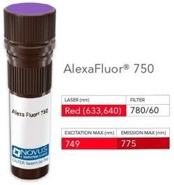
Alexa Fluor 750

Formulation

50mM Sodium Borate with 0.05% Sodium Azide

Gene Symbols

MAGEA1

Immunogen

Human MAGE 1 full length recombinant protein (Uniprot: P43355)

Quantity

0.1 mL

Research Discipline

Apoptosis, Cancer, Melanoma Cell Markers, Tumor Suppressors

Test Specificity

Recognizes a protein of 42-46kDa, identified as MAGE-1. This monoclonal antibody does not cross-react with MAGE-2, -3, -4, -6 -9, -10, -or -12 protein. Human malignant neoplasms carry rejection antigens that are recognized by the patients' autologous, tumor directed and specific, cytolytic, CD8+ T lymphocyte clones (CTL). The MAGE family of genes codes an important group of antigens. It was identified that melanomas and primary glial brain tumors express common melanoma associated antigens (MAAs). Because MAGE-1 is expressed on a significant proportion of human neoplasms of various histological types (melanoma, brain tumors of glial origin, neuroblastoma, non-small cell lung cancer, breast, gastric, colorectal, ovarian, renal cell carcinomas) and not on normal tissues, the encoded antigen may serve as a marker of early detection and target for cancer immunotherapy.

Content And Storage

Store at 4°C in the dark.

Applications

Western Blot, Flow Cytometry, ELISA, Immunohistochemistry, Immunocytochemistry, Immunofluorescence, Immunohistochemistry (Paraffin)

Clone

SPM282

Dilution

Western Blot, Flow Cytometry, ELISA, Immunohistochemistry, Immunocytochemistry/Immunofluorescence, Immunohistochemistry-Paraffin, Immunohistochemistry-Frozen

Gene Alias

Antigen MZ2-E, Cancer/testis antigen 1.1, cancer/testis antigen family 1, member 1, CT1.1melanoma antigen family A 1, MAGE-1 antigen, MAGE1A, MAGE1melanoma antigen MAGE-1, melanoma antigen family A, 1 (directs expression of antigen MZ2-E), melanoma-associated antigen 1, melanoma-associated antigen MZ2-E, MGC9326

Host Species

Mouse

Purification Method

Protein A or G purified

Regulatory Status

RUO

Primary or Secondary

Primary

Target Species

Human, Rat, Canine

Isotype

IgG1 κ

Related Products

Img

Novus Biologicals

NB006547

--

Img

Novus Biologicals

NB006548

--

Img

Novus Biologicals

NB006789

--

Img

Novus Biologicals

NB006792

--

Img

Novus Biologicals

NB006790

--

Img

Novus Biologicals

NB006726

--

Img

Novus Biologicals

NB006602

--

Img

Novus Biologicals

NB006605

--

Description

  • MAGE 1 Monoclonal specifically detects MAGE 1 in Human, Rat, Canine samples
  • It is validated for Flow Cytometry, Immunohistochemistry, Immunocytochemistry/Immunofluorescence, Immunohistochemistry-Paraffin, Flow (Intracellular).

Compare Similar Items

Show Difference

Img

Novus Biologicals

NB006550

--


Antigen:
MAGE 1

Classification:
Monoclonal

Conjugate:
Alexa Fluor 750

Formulation:
50mM Sodium Borate with 0.05% Sodium Azide

Gene Symbols:
MAGEA1

Immunogen:
Human MAGE 1 full length recombinant protein (Uniprot: P43355)

Quantity:
0.1 mL

Research Discipline:
Apoptosis, Cancer, Melanoma Cell Markers, Tumor Suppressors

Test Specificity:
Recognizes a protein of 42-46kDa, identified as MAGE-1. This monoclonal antibody does not cross-react with MAGE-2, -3, -4, -6 -9, -10, -or -12 protein. Human malignant neoplasms carry rejection antigens that are recognized by the patients' autologous, tumor directed and specific, cytolytic, CD8+ T lymphocyte clones (CTL). The MAGE family of genes codes an important group of antigens. It was identified that melanomas and primary glial brain tumors express common melanoma associated antigens (MAAs). Because MAGE-1 is expressed on a significant proportion of human neoplasms of various histological types (melanoma, brain tumors of glial origin, neuroblastoma, non-small cell lung cancer, breast, gastric, colorectal, ovarian, renal cell carcinomas) and not on normal tissues, the encoded antigen may serve as a marker of early detection and target for cancer immunotherapy.

Content And Storage:
Store at 4°C in the dark.

Applications:
Western Blot, Flow Cytometry, ELISA, Immunohistochemistry, Immunocytochemistry, Immunofluorescence, Immunohistochemistry (Paraffin)

Clone:
SPM282

Dilution:
Western Blot, Flow Cytometry, ELISA, Immunohistochemistry, Immunocytochemistry/Immunofluorescence, Immunohistochemistry-Paraffin, Immunohistochemistry-Frozen

Gene Alias:
Antigen MZ2-E, Cancer/testis antigen 1.1, cancer/testis antigen family 1, member 1, CT1.1melanoma antigen family A 1, MAGE-1 antigen, MAGE1A, MAGE1melanoma antigen MAGE-1, melanoma antigen family A, 1 (directs expression of antigen MZ2-E), melanoma-associated antigen 1, melanoma-associated antigen MZ2-E, MGC9326

Host Species:
Mouse

Purification Method:
Protein A or G purified

Regulatory Status:
RUO

Primary or Secondary:
Primary

Target Species:
Human, Rat, Canine

Isotype:
IgG1 κ

Img

Novus Biologicals

NB006553

--


Antigen:
PMEL17/SILV

Classification:
Monoclonal

Conjugate:
Alexa Fluor 350

Formulation:
50mM Sodium Borate with 0.05% Sodium Azide

Gene Symbols:
PMEL

Immunogen:
Membranes from a human melanoma metastasis

Quantity:
0.1 mL

Research Discipline:
__

Test Specificity:
By immunohistochemistry, it specifically recognizes a protein in melanocytes and melanomas. This monoclonal antibody reacts with junctional and blue nevus cells and variably with fetal and neonatal melanocytes. Intradermal nevi, normal adult melanocytes, and non-melanocytic cells are negative. It does not stain tumor cells of epithelial, lymphoid, glial, or mesenchymal origin. This Mab labels formalin-fixed, paraffin-embedded melanomas and other tumors showing melanocytic differentiation.

Content And Storage:
Store at 4°C in the dark.

Applications:
Flow Cytometry, ELISA, Immunohistochemistry, Immunocytochemistry, Immunofluorescence, Immunohistochemistry (Paraffin)

Clone:
SPM286

Dilution:
Flow Cytometry, ELISA, Immunohistochemistry, Immunocytochemistry/Immunofluorescence, Immunohistochemistry-Paraffin, Immunohistochemistry-Frozen

Gene Alias:
D12S53EP1, gp100, ME20, ME20-M, melanocyte protein mel 17, Melanocyte protein Pmel 17, Melanocytes lineage-specific antigen GP100, Melanoma-associated ME20 antigen, melanosomal matrix protein17, PMEL17P100, premelanosome proteinME20M, SI, SIL, silver (mouse homolog) like, silver homolog (mouse), Silver locus protein homolog, silver, mouse, homolog of, SILVPmel17

Host Species:
Mouse

Purification Method:
Protein A or G purified

Regulatory Status:
RUO

Primary or Secondary:
Primary

Target Species:
Human, Equine, Rat (Negative)

Isotype:
IgG2b κ

Img

Novus Biologicals

NB006554

--


Antigen:
PMEL17/SILV

Classification:
Monoclonal

Conjugate:
Alexa Fluor 532

Formulation:
50mM Sodium Borate with 0.05% Sodium Azide

Gene Symbols:
PMEL

Immunogen:
Membranes from a human melanoma metastasis

Quantity:
0.1 mL

Research Discipline:
__

Test Specificity:
By immunohistochemistry, it specifically recognizes a protein in melanocytes and melanomas. This monoclonal antibody reacts with junctional and blue nevus cells and variably with fetal and neonatal melanocytes. Intradermal nevi, normal adult melanocytes, and non-melanocytic cells are negative. It does not stain tumor cells of epithelial, lymphoid, glial, or mesenchymal origin. This Mab labels formalin-fixed, paraffin-embedded melanomas and other tumors showing melanocytic differentiation.

Content And Storage:
Store at 4°C in the dark.

Applications:
Flow Cytometry, ELISA, Immunohistochemistry, Immunocytochemistry, Immunofluorescence, Immunohistochemistry (Paraffin)

Clone:
SPM286

Dilution:
Flow Cytometry, ELISA, Immunohistochemistry, Immunocytochemistry/Immunofluorescence, Immunohistochemistry-Paraffin, Immunohistochemistry-Frozen

Gene Alias:
D12S53EP1, gp100, ME20, ME20-M, melanocyte protein mel 17, Melanocyte protein Pmel 17, Melanocytes lineage-specific antigen GP100, Melanoma-associated ME20 antigen, melanosomal matrix protein17, PMEL17P100, premelanosome proteinME20M, SI, SIL, silver (mouse homolog) like, silver homolog (mouse), Silver locus protein homolog, silver, mouse, homolog of, SILVPmel17

Host Species:
Mouse

Purification Method:
Protein A or G purified

Regulatory Status:
RUO

Primary or Secondary:
Primary

Target Species:
Human, Equine, Rat (Negative)

Isotype:
IgG2b κ

Img

Novus Biologicals

NB006555

--


Antigen:
PMEL17/SILV

Classification:
Monoclonal

Conjugate:
Alexa Fluor 594

Formulation:
50mM Sodium Borate with 0.05% Sodium Azide

Gene Symbols:
PMEL

Immunogen:
Membranes from a human melanoma metastasis

Quantity:
0.1 mL

Research Discipline:
__

Test Specificity:
By immunohistochemistry, it specifically recognizes a protein in melanocytes and melanomas. This monoclonal antibody reacts with junctional and blue nevus cells and variably with fetal and neonatal melanocytes. Intradermal nevi, normal adult melanocytes, and non-melanocytic cells are negative. It does not stain tumor cells of epithelial, lymphoid, glial, or mesenchymal origin. This Mab labels formalin-fixed, paraffin-embedded melanomas and other tumors showing melanocytic differentiation.

Content And Storage:
Store at 4°C in the dark.

Applications:
Flow Cytometry, ELISA, Immunohistochemistry, Immunocytochemistry, Immunofluorescence, Immunohistochemistry (Paraffin)

Clone:
SPM286

Dilution:
Flow Cytometry, ELISA, Immunohistochemistry, Immunocytochemistry/Immunofluorescence, Immunohistochemistry-Paraffin, Immunohistochemistry-Frozen

Gene Alias:
D12S53EP1, gp100, ME20, ME20-M, melanocyte protein mel 17, Melanocyte protein Pmel 17, Melanocytes lineage-specific antigen GP100, Melanoma-associated ME20 antigen, melanosomal matrix protein17, PMEL17P100, premelanosome proteinME20M, SI, SIL, silver (mouse homolog) like, silver homolog (mouse), Silver locus protein homolog, silver, mouse, homolog of, SILVPmel17

Host Species:
Mouse

Purification Method:
Protein A or G purified

Regulatory Status:
RUO

Primary or Secondary:
Primary

Target Species:
Human, Equine, Rat (Negative)

Isotype:
IgG2b κ