NB006736

CD5 Antibody (SPM546), Alexa Fluor™ 532, Novus Biologicals™

Manufacturer: Novus Biologicals

Select a Size

Pack Size SKU Availability Price
Each of 1 NB006736-Each-of-1 In Stock ₹ 56,555.16

NB006736 - Each of 1

₹ 56,555.16

In Stock

Quantity

1

Base Price: ₹ 56,555.16

GST (18%): ₹ 10,179.929

Total Price: ₹ 66,735.089

Antigen

CD5

Classification

Monoclonal

Conjugate

Alexa Fluor 532

Formulation

50mM Sodium Borate with 0.05% Sodium Azide

Gene Symbols

CD5

Immunogen

Human CD5 recombinant protein (Uniprot: P06127)

Quantity

0.1 mL

Research Discipline

Adaptive Immunity, B Cell Development and Differentiation Markers, Cell Biology, Cytokine Research, Immunology, Phospho Specific

Test Specificity

Recognizes a 67kDa transmembrane protein, which is identified as CD5. The CD5 antigen is found on 95% of thymocytes and 72% of peripheral blood lymphocytes. In lymph nodes, the main reactivity is observed in T cell areas. Anti-CD5 is a pan T-cell marker that also reacts with a range of neoplastic B-cells, e.g. chronic lymphocytic leukemia/small lymphocytic lymphoma (CLL/SLL), mantle cell lymphoma, and a subset (∽10%) of diffuse large B-cell lymphoma. CD5 aberrant expression is useful in making a diagnosis of mature T-cell neoplasms. Anti-CD5 detection is diagnostic in CLL/SLL within a panel of other B-cell markers, especially one that includes anti-CD23. Anti-CD5 is also very useful in differentiating among mature small lymphoid cell malignancies. In addition, anti-CD5 can be used in distinguishing thymic carcinoma (+) from thymoma (-). Anti-CD5 does not react with granulocytes or monocytes.

Content And Storage

Store at 4°C in the dark.

Applications

Western Blot, Flow Cytometry, ELISA, Immunohistochemistry, Immunocytochemistry, Immunofluorescence, Immunohistochemistry (Paraffin)

Clone

SPM546

Dilution

Western Blot, Flow Cytometry, ELISA, Immunohistochemistry, Immunocytochemistry/Immunofluorescence, Immunohistochemistry-Paraffin, Immunohistochemistry-Frozen

Gene Alias

CD5 antigen, CD5 antigen (p56-62), CD5 molecule, LEU1T-cell surface glycoprotein CD5, Lymphocyte antigen T1/Leu-1, T1

Host Species

Mouse

Purification Method

Protein A or G purified

Regulatory Status

RUO

Primary or Secondary

Primary

Target Species

Human

Isotype

IgG1 κ

Related Products

Img

Novus Biologicals

NB006737

--

Img

Novus Biologicals

NB006738

--

Img

Novus Biologicals

NB006188

--

Img

Novus Biologicals

NB006749

--

Img

Novus Biologicals

NB006747

--

Img

Novus Biologicals

NB006750

--

Img

Novus Biologicals

NB006412

--

Img

Novus Biologicals

NB006865

--

Description

  • CD5 Monoclonal specifically detects CD5 in Human samples
  • It is validated for Flow Cytometry, Immunohistochemistry, Immunocytochemistry/Immunofluorescence, Immunohistochemistry-Paraffin.
Compare Similar Items

Show Difference

Img

Novus Biologicals

NB006736

--


Antigen:
CD5

Classification:
Monoclonal

Conjugate:
Alexa Fluor 532

Formulation:
50mM Sodium Borate with 0.05% Sodium Azide

Gene Symbols:
CD5

Immunogen:
Human CD5 recombinant protein (Uniprot: P06127)

Quantity:
0.1 mL

Research Discipline:
Adaptive Immunity, B Cell Development and Differentiation Markers, Cell Biology, Cytokine Research, Immunology, Phospho Specific

Test Specificity:
Recognizes a 67kDa transmembrane protein, which is identified as CD5. The CD5 antigen is found on 95% of thymocytes and 72% of peripheral blood lymphocytes. In lymph nodes, the main reactivity is observed in T cell areas. Anti-CD5 is a pan T-cell marker that also reacts with a range of neoplastic B-cells, e.g. chronic lymphocytic leukemia/small lymphocytic lymphoma (CLL/SLL), mantle cell lymphoma, and a subset (∽10%) of diffuse large B-cell lymphoma. CD5 aberrant expression is useful in making a diagnosis of mature T-cell neoplasms. Anti-CD5 detection is diagnostic in CLL/SLL within a panel of other B-cell markers, especially one that includes anti-CD23. Anti-CD5 is also very useful in differentiating among mature small lymphoid cell malignancies. In addition, anti-CD5 can be used in distinguishing thymic carcinoma (+) from thymoma (-). Anti-CD5 does not react with granulocytes or monocytes.

Content And Storage:
Store at 4°C in the dark.

Applications:
Western Blot, Flow Cytometry, ELISA, Immunohistochemistry, Immunocytochemistry, Immunofluorescence, Immunohistochemistry (Paraffin)

Clone:
SPM546

Dilution:
Western Blot, Flow Cytometry, ELISA, Immunohistochemistry, Immunocytochemistry/Immunofluorescence, Immunohistochemistry-Paraffin, Immunohistochemistry-Frozen

Gene Alias:
CD5 antigen, CD5 antigen (p56-62), CD5 molecule, LEU1T-cell surface glycoprotein CD5, Lymphocyte antigen T1/Leu-1, T1

Host Species:
Mouse

Purification Method:
Protein A or G purified

Regulatory Status:
RUO

Primary or Secondary:
Primary

Target Species:
Human

Isotype:
IgG1 κ

Img

Novus Biologicals

NB006737

--


Antigen:
CD5

Classification:
Monoclonal

Conjugate:
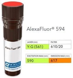
Alexa Fluor 594

Formulation:
50mM Sodium Borate with 0.05% Sodium Azide

Gene Symbols:
CD5

Immunogen:
Human CD5 recombinant protein (Uniprot: P06127)

Quantity:
0.1 mL

Research Discipline:
Adaptive Immunity, B Cell Development and Differentiation Markers, Cell Biology, Cytokine Research, Immunology, Phospho Specific

Test Specificity:
Recognizes a 67kDa transmembrane protein, which is identified as CD5. The CD5 antigen is found on 95% of thymocytes and 72% of peripheral blood lymphocytes. In lymph nodes, the main reactivity is observed in T cell areas. Anti-CD5 is a pan T-cell marker that also reacts with a range of neoplastic B-cells, e.g. chronic lymphocytic leukemia/small lymphocytic lymphoma (CLL/SLL), mantle cell lymphoma, and a subset (∽10%) of diffuse large B-cell lymphoma. CD5 aberrant expression is useful in making a diagnosis of mature T-cell neoplasms. Anti-CD5 detection is diagnostic in CLL/SLL within a panel of other B-cell markers, especially one that includes anti-CD23. Anti-CD5 is also very useful in differentiating among mature small lymphoid cell malignancies. In addition, anti-CD5 can be used in distinguishing thymic carcinoma (+) from thymoma (-). Anti-CD5 does not react with granulocytes or monocytes.

Content And Storage:
Store at 4°C in the dark.

Applications:
Western Blot, Flow Cytometry, ELISA, Immunohistochemistry, Immunocytochemistry, Immunofluorescence, Immunohistochemistry (Paraffin)

Clone:
SPM546

Dilution:
Western Blot, Flow Cytometry, ELISA, Immunohistochemistry, Immunocytochemistry/Immunofluorescence, Immunohistochemistry-Paraffin, Immunohistochemistry-Frozen

Gene Alias:
CD5 antigen, CD5 antigen (p56-62), CD5 molecule, LEU1T-cell surface glycoprotein CD5, Lymphocyte antigen T1/Leu-1, T1

Host Species:
Mouse

Purification Method:
Protein A or G purified

Regulatory Status:
RUO

Primary or Secondary:
Primary

Target Species:
Human

Isotype:
IgG1 κ

Img

Novus Biologicals

NB006738

--


Antigen:
CD5

Classification:
Monoclonal

Conjugate:
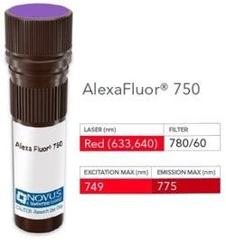
Alexa Fluor 750

Formulation:
50mM Sodium Borate with 0.05% Sodium Azide

Gene Symbols:
CD5

Immunogen:
Human CD5 recombinant protein (Uniprot: P06127)

Quantity:
0.1 mL

Research Discipline:
Adaptive Immunity, B Cell Development and Differentiation Markers, Cell Biology, Cytokine Research, Immunology, Phospho Specific

Test Specificity:
Recognizes a 67kDa transmembrane protein, which is identified as CD5. The CD5 antigen is found on 95% of thymocytes and 72% of peripheral blood lymphocytes. In lymph nodes, the main reactivity is observed in T cell areas. Anti-CD5 is a pan T-cell marker that also reacts with a range of neoplastic B-cells, e.g. chronic lymphocytic leukemia/small lymphocytic lymphoma (CLL/SLL), mantle cell lymphoma, and a subset (∽10%) of diffuse large B-cell lymphoma. CD5 aberrant expression is useful in making a diagnosis of mature T-cell neoplasms. Anti-CD5 detection is diagnostic in CLL/SLL within a panel of other B-cell markers, especially one that includes anti-CD23. Anti-CD5 is also very useful in differentiating among mature small lymphoid cell malignancies. In addition, anti-CD5 can be used in distinguishing thymic carcinoma (+) from thymoma (-). Anti-CD5 does not react with granulocytes or monocytes.

Content And Storage:
Store at 4°C in the dark.

Applications:
Western Blot, Flow Cytometry, ELISA, Immunohistochemistry, Immunocytochemistry, Immunofluorescence, Immunohistochemistry (Paraffin)

Clone:
SPM546

Dilution:
Western Blot, Flow Cytometry, ELISA, Immunohistochemistry, Immunocytochemistry/Immunofluorescence, Immunohistochemistry-Paraffin, Immunohistochemistry-Frozen

Gene Alias:
CD5 antigen, CD5 antigen (p56-62), CD5 molecule, LEU1T-cell surface glycoprotein CD5, Lymphocyte antigen T1/Leu-1, T1

Host Species:
Mouse

Purification Method:
Protein A or G purified

Regulatory Status:
RUO

Primary or Secondary:
Primary

Target Species:
Human

Isotype:
IgG1 κ

Img

Novus Biologicals

NB006743

--


Antigen:
CD6

Classification:
Monoclonal

Conjugate:
Alexa Fluor 594

Formulation:
50mM Sodium Borate with 0.05% Sodium Azide

Gene Symbols:
CD6

Immunogen:
Human recombinant CD6 protein (Uniprot: P30203)

Quantity:
0.1 mL

Research Discipline:
Cytokine Research, Immunology, Signal Transduction

Test Specificity:
CD6 is a type I transmembrane glycoprotein that contains a 24-amino acid signal sequence, three extracellular scavenger receptor cysteine-rich (SRCR) domains, a membrane-spanning domain and a 44-amino acid cytoplasmic domain. The CD6 glycoprotein is tyrosine phosphorylated during TCR-mediated T cell activation. CD6 shows significant homology to CD5. CD6 is present on mature thymocytes, peripheral T cells and a subset of B cells. Antibodies to CD6 are used to deplete T cells from bone marrow transplants to prevent graft versus host disease.

Content And Storage:
Store at 4°C in the dark.

Applications:
Western Blot, ELISA, Immunohistochemistry, Immunocytochemistry, Immunofluorescence, Immunohistochemistry (Paraffin)

Clone:
SPM547

Dilution:
Western Blot, ELISA, Immunohistochemistry, Immunocytochemistry/Immunofluorescence, Immunohistochemistry-Paraffin, Immunohistochemistry-Frozen

Gene Alias:
CD6 antigenFLJ44171, CD6 molecule, T12, Tp120, TP120T-cell differentiation antigen CD6

Host Species:
Mouse

Purification Method:
Protein A or G purified

Regulatory Status:
RUO

Primary or Secondary:
Primary

Target Species:
Human

Isotype:
IgG1 κ