NF-H Antibody (RT-97 + NR-4), DyLight 405, Novus Biologicals™
Manufacturer: Novus Biologicals
Select a Size
| Pack Size | SKU | Availability | Price |
|---|---|---|---|
| Each of 1 | NB007110-Each-of-1 | In Stock | ₹ 57,494.00 |
NB007110 - Each of 1
In Stock
Quantity
1
Base Price: ₹ 57,494.00
GST (18%): ₹ 10,348.92
Total Price: ₹ 67,842.92
Antigen
NF-H
Classification
Monoclonal
Conjugate
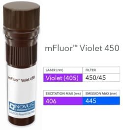
DyLight 405
Formulation
50mM Sodium Borate with 0.05% Sodium Azide
Gene Symbols
NEFH
Immunogen
Triton-X 100 insoluble proteins of rat brain (RT-97); Neurofilaments from porcine spinal cord (NR-4) (Uniprot: P12036)
Quantity
0.1 mL
Research Discipline
Autophagy, Cell Biology, Cellular Markers, Neurodegeneration, Neuronal Cell Markers, Neuroscience
Test Specificity
This monoclonal antibody reacts with a 200kDa and 68kDa protein, identified as heavy and light sub-units of neurofilaments (NF-H NF-L). Neurofilaments make up the main structural elements of axons and dendrites and are found in neurons, peripheral nerves, and sympathetic ganglion cells. Neurofilaments consist of three major subunits with molecular weights of 68kDa (NF-L), 160kDa (NF-M) and 200kDa (NF-H). Anti-neurofilament stains a number of neural, neuroendocrine, and endocrine tumors. Neuromas, ganglioneuromas, gangliogliomas, ganglioneuroblastomas, and neuroblastomas stain positively for anti-neurofilament. Neurofilaments are also present in paragangliomas as well as adrenal and extra-adrenal pheochromocytomas. Carcinoids, neuroendocrine carcinomas of the skin, and oat cell carcinomas of the lung also express neurofilament.
Content And Storage
Store at 4°C in the dark.
Applications
Flow Cytometry, Immunohistochemistry, Immunohistochemistry (Paraffin)
Clone
RT-97 + NR-4
Dilution
Flow Cytometry, Immunohistochemistry, Immunohistochemistry-Paraffin
Gene Alias
200 kDa neurofilament protein, KIAA0845, Neurofilament Heavy (200kDa), neurofilament heavy polypeptide, Neurofilament triplet H protein, neurofilament, heavy polypeptide, neurofilament, heavy polypeptide 200kDa, NF200, NFH, NF-H
Host Species
Mouse
Purification Method
Protein G purified
Regulatory Status
RUO
Primary or Secondary
Primary
Target Species
Human, Mouse, Rat, Porcine, Chicken
Isotype
IgG1 κ
Related Products


Description
- NF-H Monoclonal specifically detects NF-H in Human, Mouse, Rat, Porcine, Chicken samples
- It is validated for Flow Cytometry, Immunohistochemistry, Immunohistochemistry-Paraffin.
Compare Similar Items
Show Difference
Antigen: NF-H
Classification: Monoclonal
Conjugate: DyLight 405
Formulation: 50mM Sodium Borate with 0.05% Sodium Azide
Gene Symbols: NEFH
Immunogen: Triton-X 100 insoluble proteins of rat brain (RT-97); Neurofilaments from porcine spinal cord (NR-4) (Uniprot: P12036)
Quantity: 0.1 mL
Research Discipline: Autophagy, Cell Biology, Cellular Markers, Neurodegeneration, Neuronal Cell Markers, Neuroscience
Test Specificity: This monoclonal antibody reacts with a 200kDa and 68kDa protein, identified as heavy and light sub-units of neurofilaments (NF-H NF-L). Neurofilaments make up the main structural elements of axons and dendrites and are found in neurons, peripheral nerves, and sympathetic ganglion cells. Neurofilaments consist of three major subunits with molecular weights of 68kDa (NF-L), 160kDa (NF-M) and 200kDa (NF-H). Anti-neurofilament stains a number of neural, neuroendocrine, and endocrine tumors. Neuromas, ganglioneuromas, gangliogliomas, ganglioneuroblastomas, and neuroblastomas stain positively for anti-neurofilament. Neurofilaments are also present in paragangliomas as well as adrenal and extra-adrenal pheochromocytomas. Carcinoids, neuroendocrine carcinomas of the skin, and oat cell carcinomas of the lung also express neurofilament.
Content And Storage: Store at 4°C in the dark.
Applications: Flow Cytometry, Immunohistochemistry, Immunohistochemistry (Paraffin)
Clone: RT-97 + NR-4
Dilution: Flow Cytometry, Immunohistochemistry, Immunohistochemistry-Paraffin
Gene Alias: 200 kDa neurofilament protein, KIAA0845, Neurofilament Heavy (200kDa), neurofilament heavy polypeptide, Neurofilament triplet H protein, neurofilament, heavy polypeptide, neurofilament, heavy polypeptide 200kDa, NF200, NFH, NF-H
Host Species: Mouse
Purification Method: Protein G purified
Regulatory Status: RUO
Primary or Secondary: Primary
Target Species: Human, Mouse, Rat, Porcine, Chicken
Isotype: IgG1 κ

Antigen: S100A8/A9
Classification: Monoclonal
Conjugate: Alexa Fluor 350
Formulation: 50mM Sodium Borate with 0.05% Sodium Azide
Gene Symbols: S100A8
Immunogen: Affinity purified monocyte membrane preparation
Quantity: 0.1 mL
Research Discipline: Cancer
Test Specificity: Recognizes the L1 or Calprotectin molecule, an intra-cytoplasmic antigen comprising of a 12kDa alpha chain and a 14kDa beta chain expressed by granulocytes, monocytes and by tissue macrophages. Macrophages usually arise from hematopoietic stem cells in the bone marrow. Under migration into tissues, the monocytes undergo further differentiation to become multifunctional tissue macrophages. They are classified into normal and inflammatory macrophages. Normal macrophages include macrophages in connective tissue (histiocytes), liver (Kupffers cell) and in other tissues. Inflammatory macrophages are present in various exudates. Macrophages are part of the innate immune system, recognizing, engulfing and destroying many potential pathogens including bacteria, pathogenic protozoa, fungi and helminthes. This monoclonal antibody reacts with neutrophils, monocytes, macrophages, and squamous mucosal epithelia and has been shown as an important marker for identifying macrophages in tissue sections.
Content And Storage: Store at 4°C in the dark.
Applications: Flow Cytometry, Immunohistochemistry, Immunohistochemistry (Paraffin), Immunofluorescence
Clone: MAC387
Dilution: Flow Cytometry, Immunohistochemistry, Immunohistochemistry-Paraffin, Immunofluorescence
Gene Alias: 60B8AG, CAGA, CFAG, CGLA, CP-10, L1Ag, MA387, MIF, MRP8, NIF, P8, S100 calcium binding protein A8, S100A8
Host Species: Mouse
Purification Method: Protein A or G purified
Regulatory Status: RUO
Primary or Secondary: Primary
Target Species: Human, Mouse, Rat, Porcine, Canine, Equine, Feline, Guinea Pig, Goat, Baboon, Monkey, Rabbit
Isotype: IgG1 κ

Antigen: S100A8/A9
Classification: Monoclonal
Conjugate: Alexa Fluor 750
Formulation: 50mM Sodium Borate with 0.05% Sodium Azide
Gene Symbols: S100A8
Immunogen: Affinity purified monocyte membrane preparation
Quantity: 0.1 mL
Research Discipline: Cancer
Test Specificity: Recognizes the L1 or Calprotectin molecule, an intra-cytoplasmic antigen comprising of a 12kDa alpha chain and a 14kDa beta chain expressed by granulocytes, monocytes and by tissue macrophages. Macrophages usually arise from hematopoietic stem cells in the bone marrow. Under migration into tissues, the monocytes undergo further differentiation to become multifunctional tissue macrophages. They are classified into normal and inflammatory macrophages. Normal macrophages include macrophages in connective tissue (histiocytes), liver (Kupffers cell) and in other tissues. Inflammatory macrophages are present in various exudates. Macrophages are part of the innate immune system, recognizing, engulfing and destroying many potential pathogens including bacteria, pathogenic protozoa, fungi and helminthes. This monoclonal antibody reacts with neutrophils, monocytes, macrophages, and squamous mucosal epithelia and has been shown as an important marker for identifying macrophages in tissue sections.
Content And Storage: Store at 4°C in the dark.
Applications: Flow Cytometry, Immunohistochemistry, Immunohistochemistry (Paraffin), Immunofluorescence
Clone: MAC387
Dilution: Flow Cytometry, Immunohistochemistry, Immunohistochemistry-Paraffin, Immunofluorescence
Gene Alias: 60B8AG, CAGA, CFAG, CGLA, CP-10, L1Ag, MA387, MIF, MRP8, NIF, P8, S100 calcium binding protein A8, S100A8
Host Species: Mouse
Purification Method: Protein A or G purified
Regulatory Status: RUO
Primary or Secondary: Primary
Target Species: Human, Mouse, Rat, Porcine, Canine, Equine, Feline, Guinea Pig, Goat, Baboon, Monkey, Rabbit
Isotype: IgG1 κ