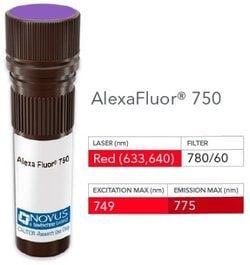
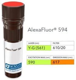
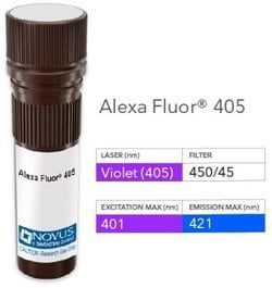
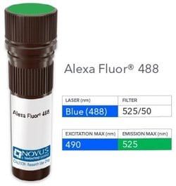
NB007207

LH beta Antibody (LHb/1214), DyLight 594, Novus Biologicals™

Manufacturer: Novus Biologicals

Select a Size

Pack Size SKU Availability Price
Each of 1 NB007207-Each-of-1 In Stock ₹ 55,271.76

NB007207 - Each of 1

₹ 55,271.76

In Stock

Quantity

1

Base Price: ₹ 55,271.76

GST (18%): ₹ 9,948.917

Total Price: ₹ 65,220.677

Antigen

LH beta

Classification

Monoclonal

Conjugate

DyLight 594

Formulation

50mM Sodium Borate with 0.05% Sodium Azide

Gene Symbols

LHB

Immunogen

Recombinant beta sub-unit of human LH beta (Uniprot: P01229)

Quantity

0.1 mL

Primary or Secondary

Primary

Target Species

Human

Isotype

IgG1 κ

Applications

Flow Cytometry, Immunohistochemistry, Immunohistochemistry (Paraffin), Immunofluorescence

Clone

LHb/1214

Dilution

Flow Cytometry, Immunohistochemistry, Immunohistochemistry-Paraffin, Immunofluorescence

Gene Alias

CGB4, hLHB, interstitial cell stimulating hormone, beta chain, LSH-B, LSH-beta, luteinizing hormone beta polypeptide, luteinizing hormone beta subunit, lutropin beta chain, lutropin subunit beta

Host Species

Mouse

Purification Method

Protein A or G purified

Regulatory Status

RUO

Test Specificity

Luteinizing hormone (LH) is a glycoprotein. Each monomeric unit is a sugar-like protein molecule; two of these make the full, functional protein. Its structure is similar to the other glycoproteins, follicle-stimulating hormone (FSH), thyroid-stimulating hormone (TSH), and human chorionic gonadotropin (hCG). The protein dimer contains 2 polypeptide units, labeled alpha and beta subunits that are connected by two bridges. The alpha subunits of LH, FSH, TSH, and hCG are identical, and contain 92 amino acids. The beta subunits vary. LH has a beta subunit of 121 amino acids (LHB) that confers its specific biologic action and is responsible for interaction with the LH receptor. This beta subunit contains the same amino acids in sequence as the beta subunit of hCG and both stimulate the same receptor; however, the hCG beta subunit contains an additional 24 amino acids and the hormones differ in the composition of their sugar moieties.LH is synthesized and secreted by gonadotrophs in the anterior lobe of the pituitary gland. In concert with the other pituitary gonadotropin follicle-stimulating hormone (FSH), it is necessary for proper reproductive function. In the female, an acute rise of LH levels triggers ovulation. In the male, where LH has also been called Interstitial Cell-Stimulating Hormone (ICSH), it stimulates Leydig cell production of testosterone. LH is a useful marker in classification of pituitary tumors and the study of pituitary disease.

Content And Storage

Store at 4°C in the dark.

Related Products

Img

Novus Biologicals

NB005802

--

Img

Novus Biologicals

NB005939

--

Img

Novus Biologicals

NB005940

--

Img

Novus Biologicals

NB005942

--

Img

Novus Biologicals

NB005801

--

Img

Novus Biologicals

NB006266

--

Img

Novus Biologicals

NB005803

--

Img

Novus Biologicals

NB006591

--

Description

  • LH beta Monoclonal specifically detects LH beta in Human samples
  • It is validated for Immunohistochemistry, Immunohistochemistry-Paraffin.

Compare Similar Items

Show Difference

Img

Novus Biologicals

NB007207

--


Antigen:
LH beta

Classification:
Monoclonal

Conjugate:
DyLight 594

Formulation:
50mM Sodium Borate with 0.05% Sodium Azide

Gene Symbols:
LHB

Immunogen:
Recombinant beta sub-unit of human LH beta (Uniprot: P01229)

Quantity:
0.1 mL

Primary or Secondary:
Primary

Target Species:
Human

Isotype:
IgG1 κ

Applications:
Flow Cytometry, Immunohistochemistry, Immunohistochemistry (Paraffin), Immunofluorescence

Clone:
LHb/1214

Dilution:
Flow Cytometry, Immunohistochemistry, Immunohistochemistry-Paraffin, Immunofluorescence

Gene Alias:
CGB4, hLHB, interstitial cell stimulating hormone, beta chain, LSH-B, LSH-beta, luteinizing hormone beta polypeptide, luteinizing hormone beta subunit, lutropin beta chain, lutropin subunit beta

Host Species:
Mouse

Purification Method:
Protein A or G purified

Regulatory Status:
RUO

Test Specificity:
Luteinizing hormone (LH) is a glycoprotein. Each monomeric unit is a sugar-like protein molecule; two of these make the full, functional protein. Its structure is similar to the other glycoproteins, follicle-stimulating hormone (FSH), thyroid-stimulating hormone (TSH), and human chorionic gonadotropin (hCG). The protein dimer contains 2 polypeptide units, labeled alpha and beta subunits that are connected by two bridges. The alpha subunits of LH, FSH, TSH, and hCG are identical, and contain 92 amino acids. The beta subunits vary. LH has a beta subunit of 121 amino acids (LHB) that confers its specific biologic action and is responsible for interaction with the LH receptor. This beta subunit contains the same amino acids in sequence as the beta subunit of hCG and both stimulate the same receptor; however, the hCG beta subunit contains an additional 24 amino acids and the hormones differ in the composition of their sugar moieties.LH is synthesized and secreted by gonadotrophs in the anterior lobe of the pituitary gland. In concert with the other pituitary gonadotropin follicle-stimulating hormone (FSH), it is necessary for proper reproductive function. In the female, an acute rise of LH levels triggers ovulation. In the male, where LH has also been called Interstitial Cell-Stimulating Hormone (ICSH), it stimulates Leydig cell production of testosterone. LH is a useful marker in classification of pituitary tumors and the study of pituitary disease.

Content And Storage:
Store at 4°C in the dark.

Img

Novus Biologicals

NB007208

--


Antigen:
EGFR

Classification:
Monoclonal

Conjugate:
Alexa Fluor 350

Formulation:
50mM Sodium Borate with 0.05% Sodium Azide

Gene Symbols:
EGFR

Immunogen:
Human EGFR purified from A431 cells (31G7); Recombinant human EGFR protein (GFR1195)

Quantity:
0.1 mL

Primary or Secondary:
Primary

Target Species:
Human

Isotype:
IgG2a κ

Applications:
Flow Cytometry, Immunohistochemistry, Immunohistochemistry (Paraffin), Immunofluorescence

Clone:
31G7 + GFR1195

Dilution:
Flow Cytometry, Immunohistochemistry, Immunohistochemistry-Paraffin, Flow (Cell Surface), Immunofluorescence

Gene Alias:
avian erythroblastic leukemia viral (v-erb-b) oncogene homolog, cell growth inhibiting protein 40, cell proliferation-inducing protein 61, EC 2.7.10, EC 2.7.10.1, epidermal growth factor receptor, epidermal growth factor receptor (avian erythroblastic leukemia viral (v-erb-b)oncogene homolog), ERBB, ErbB1, ERBB1PIG61, HER1, mENA, Proto-oncogene c-ErbB-1, Receptor tyrosine-protein kinase erbB-1

Host Species:
Mouse

Purification Method:
Protein A or G purified

Regulatory Status:
RUO

Test Specificity:
This MAb recognizes a protein of 170kDa, identified as EGFR. EGFR is type I receptor tyrosine kinase with sequence homology to erbB-1, -2, -3 -4 or HER-1, -2, -3 -4. It binds to Epidermal Growth Factor (EGF), Transforming Growth Factor-a (TGF-a), Heparin-binding EGF (HB-EGF), amphiregulin, Beta cellulin and epiregulin. EGFR is overexpressed in tumors of breast, brain, bladder, lung, gastric, head & neck, esophagus, cervix, vulva, ovary, and endometrium. It is predominantly present in squamous cell carcinomas.

Content And Storage:
Store at 4°C in the dark.

Img

Novus Biologicals

NB007209

--


Antigen:
EGFR

Classification:
Monoclonal

Conjugate:
Alexa Fluor 405

Formulation:
50mM Sodium Borate with 0.05% Sodium Azide

Gene Symbols:
EGFR

Immunogen:
Human EGFR purified from A431 cells (31G7); Recombinant human EGFR protein (GFR1195)

Quantity:
0.1 mL

Primary or Secondary:
Primary

Target Species:
Human

Isotype:
IgG2a κ

Applications:
Flow Cytometry, Immunohistochemistry, Immunohistochemistry (Paraffin), Immunofluorescence

Clone:
31G7 + GFR1195

Dilution:
Flow Cytometry, Immunohistochemistry, Immunohistochemistry-Paraffin, Flow (Cell Surface), Immunofluorescence

Gene Alias:
avian erythroblastic leukemia viral (v-erb-b) oncogene homolog, cell growth inhibiting protein 40, cell proliferation-inducing protein 61, EC 2.7.10, EC 2.7.10.1, epidermal growth factor receptor, epidermal growth factor receptor (avian erythroblastic leukemia viral (v-erb-b)oncogene homolog), ERBB, ErbB1, ERBB1PIG61, HER1, mENA, Proto-oncogene c-ErbB-1, Receptor tyrosine-protein kinase erbB-1

Host Species:
Mouse

Purification Method:
Protein A or G purified

Regulatory Status:
RUO

Test Specificity:
This MAb recognizes a protein of 170kDa, identified as EGFR. EGFR is type I receptor tyrosine kinase with sequence homology to erbB-1, -2, -3 -4 or HER-1, -2, -3 -4. It binds to Epidermal Growth Factor (EGF), Transforming Growth Factor-a (TGF-a), Heparin-binding EGF (HB-EGF), amphiregulin, Beta cellulin and epiregulin. EGFR is overexpressed in tumors of breast, brain, bladder, lung, gastric, head & neck, esophagus, cervix, vulva, ovary, and endometrium. It is predominantly present in squamous cell carcinomas.

Content And Storage:
Store at 4°C in the dark.

Img

Novus Biologicals

NB007210

--


Antigen:
EGFR

Classification:
Monoclonal

Conjugate:
Alexa Fluor 488

Formulation:
50mM Sodium Borate with 0.05% Sodium Azide

Gene Symbols:
EGFR

Immunogen:
Human EGFR purified from A431 cells (31G7); Recombinant human EGFR protein (GFR1195)

Quantity:
0.1 mL

Primary or Secondary:
Primary

Target Species:
Human

Isotype:
IgG2a κ

Applications:
Flow Cytometry, Immunohistochemistry, Immunohistochemistry (Paraffin), Immunofluorescence

Clone:
31G7 + GFR1195

Dilution:
Flow Cytometry, Immunohistochemistry, Immunohistochemistry-Paraffin, Flow (Cell Surface), Immunofluorescence

Gene Alias:
avian erythroblastic leukemia viral (v-erb-b) oncogene homolog, cell growth inhibiting protein 40, cell proliferation-inducing protein 61, EC 2.7.10, EC 2.7.10.1, epidermal growth factor receptor, epidermal growth factor receptor (avian erythroblastic leukemia viral (v-erb-b)oncogene homolog), ERBB, ErbB1, ERBB1PIG61, HER1, mENA, Proto-oncogene c-ErbB-1, Receptor tyrosine-protein kinase erbB-1

Host Species:
Mouse

Purification Method:
Protein A or G purified

Regulatory Status:
RUO

Test Specificity:
This MAb recognizes a protein of 170kDa, identified as EGFR. EGFR is type I receptor tyrosine kinase with sequence homology to erbB-1, -2, -3 -4 or HER-1, -2, -3 -4. It binds to Epidermal Growth Factor (EGF), Transforming Growth Factor-a (TGF-a), Heparin-binding EGF (HB-EGF), amphiregulin, Beta cellulin and epiregulin. EGFR is overexpressed in tumors of breast, brain, bladder, lung, gastric, head & neck, esophagus, cervix, vulva, ovary, and endometrium. It is predominantly present in squamous cell carcinomas.

Content And Storage:
Store at 4°C in the dark.