NB007250

H1F0 Antibody (AE-4), Alexa Fluor™ 350, Novus Biologicals™

Manufacturer: Novus Biologicals

Select a Size

Pack Size SKU Availability Price
Each of 1 NB007250-Each-of-1 In Stock ₹ 55,271.76

NB007250 - Each of 1

₹ 55,271.76

In Stock

Quantity

1

Base Price: ₹ 55,271.76

GST (18%): ₹ 9,948.917

Total Price: ₹ 65,220.677

Antigen

H1F0

Classification

Monoclonal

Conjugate

Alexa Fluor 350

Formulation

50mM Sodium Borate with 0.05% Sodium Azide

Gene Symbols

H1-0

Immunogen

Nuclei of human leukemia biopsy cells

Quantity

0.1 mL

Research Discipline

Cellular Markers, Chromatin Research, Epigenetics, Nuclear Envelope Markers

Gene ID (Entrez)

3005

Target Species

Human, Mouse, Rat

Isotype

IgG2a κ

Applications

Flow Cytometry, Immunohistochemistry, Immunohistochemistry (Paraffin), Immunofluorescence

Clone

AE-4

Dilution

Flow Cytometry, Immunohistochemistry, Immunohistochemistry-Paraffin, Immunofluorescence

Gene Alias

H1 histone family, member 0, H1.0, H1(0), H1-0, H10, H1FVMGC5241, Histone H1', Histone H1(0), histone H1.0

Host Species

Mouse

Purification Method

Protein A or G purified

Regulatory Status

RUO

Primary or Secondary

Primary

Test Specificity

Eukaryotic histones are basic and water-soluble nuclear proteins that form hetero-octameric nucleosome particles by wrapping 146 base pairs of DNA in a left-handed super-helical turn sequentially to form chromosomal fiber. Two molecules of each of the four core histones (H2A, H2B, H3, and H4) form the octamer; formed of two H2A-H2B dimers and two H3-H4 dimers, forming two nearly symmetrical halves by tertiary structure. Over 80% of nucleosomes contain the linker Histone H1, derived from an intronless gene that interacts with linker DNA between nucleosomes and mediates compaction into higher order chromatin. Histones are subject to posttranslational modification by enzymes primarily on their N-terminal tails, but also in their globular domains. Such modifications include methylation, citrullination, acetylation, phosphorylation, sumoylation, ubiquitination and ADP-ribosylation.

Content And Storage

Store at 4°C in the dark.

Related Products

Img

Novus Biologicals

NB006117

--

Img

Novus Biologicals

NB006244

--

Img

Novus Biologicals

NB11039113C

--

Img

Novus Biologicals

N234432J646

--

Img

Novus Biologicals

NB007285

--

Img

Novus Biologicals

NB007301

--

Img

Novus Biologicals

NB009224

--

Img

Novus Biologicals

NB009245

--

Description

  • H1F0 Monoclonal antibody specifically detects H1F0 in Human, Mouse, Rat samples
  • It is validated for Flow Cytometry, Immunohistochemistry, Immunocytochemistry, Immunofluorescence, Immunohistochemistry (Paraffin).

Compare Similar Items

Show Difference

Img

Novus Biologicals

NB007250

--


Antigen:
H1F0

Classification:
Monoclonal

Conjugate:
Alexa Fluor 350

Formulation:
50mM Sodium Borate with 0.05% Sodium Azide

Gene Symbols:
H1-0

Immunogen:
Nuclei of human leukemia biopsy cells

Quantity:
0.1 mL

Research Discipline:
Cellular Markers, Chromatin Research, Epigenetics, Nuclear Envelope Markers

Gene ID (Entrez):
3005

Target Species:
Human, Mouse, Rat

Isotype:
IgG2a κ

Applications:
Flow Cytometry, Immunohistochemistry, Immunohistochemistry (Paraffin), Immunofluorescence

Clone:
AE-4

Dilution:
Flow Cytometry, Immunohistochemistry, Immunohistochemistry-Paraffin, Immunofluorescence

Gene Alias:
H1 histone family, member 0, H1.0, H1(0), H1-0, H10, H1FVMGC5241, Histone H1', Histone H1(0), histone H1.0

Host Species:
Mouse

Purification Method:
Protein A or G purified

Regulatory Status:
RUO

Primary or Secondary:
Primary

Test Specificity:
Eukaryotic histones are basic and water-soluble nuclear proteins that form hetero-octameric nucleosome particles by wrapping 146 base pairs of DNA in a left-handed super-helical turn sequentially to form chromosomal fiber. Two molecules of each of the four core histones (H2A, H2B, H3, and H4) form the octamer; formed of two H2A-H2B dimers and two H3-H4 dimers, forming two nearly symmetrical halves by tertiary structure. Over 80% of nucleosomes contain the linker Histone H1, derived from an intronless gene that interacts with linker DNA between nucleosomes and mediates compaction into higher order chromatin. Histones are subject to posttranslational modification by enzymes primarily on their N-terminal tails, but also in their globular domains. Such modifications include methylation, citrullination, acetylation, phosphorylation, sumoylation, ubiquitination and ADP-ribosylation.

Content And Storage:
Store at 4°C in the dark.

Img

Novus Biologicals

NB007251

--


Antigen:
H1F0

Classification:
Monoclonal

Conjugate:
Alexa Fluor 532

Formulation:
50mM Sodium Borate with 0.05% Sodium Azide

Gene Symbols:
H1-0

Immunogen:
Nuclei of human leukemia biopsy cells

Quantity:
0.1 mL

Research Discipline:
Cellular Markers, Chromatin Research, Epigenetics, Nuclear Envelope Markers

Gene ID (Entrez):
3005

Target Species:
Human, Mouse, Rat

Isotype:
IgG2a κ

Applications:
Flow Cytometry, Immunohistochemistry, Immunohistochemistry (Paraffin), Immunofluorescence

Clone:
AE-4

Dilution:
Flow Cytometry, Immunohistochemistry, Immunohistochemistry-Paraffin, Immunofluorescence

Gene Alias:
H1 histone family, member 0, H1.0, H1(0), H1-0, H10, H1FVMGC5241, Histone H1', Histone H1(0), histone H1.0

Host Species:
Mouse

Purification Method:
Protein A or G purified

Regulatory Status:
RUO

Primary or Secondary:
Primary

Test Specificity:
Eukaryotic histones are basic and water-soluble nuclear proteins that form hetero-octameric nucleosome particles by wrapping 146 base pairs of DNA in a left-handed super-helical turn sequentially to form chromosomal fiber. Two molecules of each of the four core histones (H2A, H2B, H3, and H4) form the octamer; formed of two H2A-H2B dimers and two H3-H4 dimers, forming two nearly symmetrical halves by tertiary structure. Over 80% of nucleosomes contain the linker Histone H1, derived from an intronless gene that interacts with linker DNA between nucleosomes and mediates compaction into higher order chromatin. Histones are subject to posttranslational modification by enzymes primarily on their N-terminal tails, but also in their globular domains. Such modifications include methylation, citrullination, acetylation, phosphorylation, sumoylation, ubiquitination and ADP-ribosylation.

Content And Storage:
Store at 4°C in the dark.

Img

Novus Biologicals

NB007252

--


Antigen:
H1F0

Classification:
Monoclonal

Conjugate:
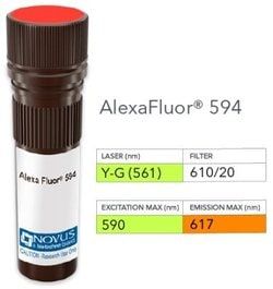
Alexa Fluor 594

Formulation:
50mM Sodium Borate with 0.05% Sodium Azide

Gene Symbols:
H1-0

Immunogen:
Nuclei of human leukemia biopsy cells

Quantity:
0.1 mL

Research Discipline:
Cellular Markers, Chromatin Research, Epigenetics, Nuclear Envelope Markers

Gene ID (Entrez):
3005

Target Species:
Human, Mouse, Rat

Isotype:
IgG2a κ

Applications:
Flow Cytometry, Immunohistochemistry, Immunohistochemistry (Paraffin), Immunofluorescence

Clone:
AE-4

Dilution:
Flow Cytometry, Immunohistochemistry, Immunohistochemistry-Paraffin, Immunofluorescence

Gene Alias:
H1 histone family, member 0, H1.0, H1(0), H1-0, H10, H1FVMGC5241, Histone H1', Histone H1(0), histone H1.0

Host Species:
Mouse

Purification Method:
Protein A or G purified

Regulatory Status:
RUO

Primary or Secondary:
Primary

Test Specificity:
Eukaryotic histones are basic and water-soluble nuclear proteins that form hetero-octameric nucleosome particles by wrapping 146 base pairs of DNA in a left-handed super-helical turn sequentially to form chromosomal fiber. Two molecules of each of the four core histones (H2A, H2B, H3, and H4) form the octamer; formed of two H2A-H2B dimers and two H3-H4 dimers, forming two nearly symmetrical halves by tertiary structure. Over 80% of nucleosomes contain the linker Histone H1, derived from an intronless gene that interacts with linker DNA between nucleosomes and mediates compaction into higher order chromatin. Histones are subject to posttranslational modification by enzymes primarily on their N-terminal tails, but also in their globular domains. Such modifications include methylation, citrullination, acetylation, phosphorylation, sumoylation, ubiquitination and ADP-ribosylation.

Content And Storage:
Store at 4°C in the dark.

Img

Novus Biologicals

NB007253

--


Antigen:
H1F0

Classification:
Monoclonal

Conjugate:
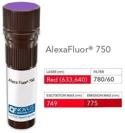
Alexa Fluor 750

Formulation:
50mM Sodium Borate with 0.05% Sodium Azide

Gene Symbols:
H1-0

Immunogen:
Nuclei of human leukemia biopsy cells

Quantity:
0.1 mL

Research Discipline:
Cellular Markers, Chromatin Research, Epigenetics, Nuclear Envelope Markers

Gene ID (Entrez):
3005

Target Species:
Human, Mouse, Rat

Isotype:
IgG2a κ

Applications:
Flow Cytometry, Immunohistochemistry, Immunohistochemistry (Paraffin), Immunofluorescence

Clone:
AE-4

Dilution:
Flow Cytometry, Immunohistochemistry, Immunohistochemistry-Paraffin, Immunofluorescence

Gene Alias:
H1 histone family, member 0, H1.0, H1(0), H1-0, H10, H1FVMGC5241, Histone H1', Histone H1(0), histone H1.0

Host Species:
Mouse

Purification Method:
Protein A or G purified

Regulatory Status:
RUO

Primary or Secondary:
Primary

Test Specificity:
Eukaryotic histones are basic and water-soluble nuclear proteins that form hetero-octameric nucleosome particles by wrapping 146 base pairs of DNA in a left-handed super-helical turn sequentially to form chromosomal fiber. Two molecules of each of the four core histones (H2A, H2B, H3, and H4) form the octamer; formed of two H2A-H2B dimers and two H3-H4 dimers, forming two nearly symmetrical halves by tertiary structure. Over 80% of nucleosomes contain the linker Histone H1, derived from an intronless gene that interacts with linker DNA between nucleosomes and mediates compaction into higher order chromatin. Histones are subject to posttranslational modification by enzymes primarily on their N-terminal tails, but also in their globular domains. Such modifications include methylation, citrullination, acetylation, phosphorylation, sumoylation, ubiquitination and ADP-ribosylation.

Content And Storage:
Store at 4°C in the dark.