NB007350

ZAP70 Antibody (ZAP70/528 + 2F3.2), DyLight 594, Novus Biologicals™

Manufacturer: Novus Biologicals

Select a Size

Pack Size SKU Availability Price
Each of 1 NB007350-Each-of-1 In Stock ₹ 55,271.76

NB007350 - Each of 1

₹ 55,271.76

In Stock

Quantity

1

Base Price: ₹ 55,271.76

GST (18%): ₹ 9,948.917

Total Price: ₹ 65,220.677

Antigen

ZAP70

Classification

Monoclonal

Conjugate

DyLight 594

Formulation

50mM Sodium Borate with 0.05% Sodium Azide

Gene Symbols

ZAP70

Immunogen

Recombinant full-length human ZAP70 protein (ZAP70/528); Recombinant ZAP-70 protein including residues 1-254 and encompassing SH2 domains of human ZAP70 (2F3.2) (Uniprot: P43403)

Quantity

0.1 mL

Research Discipline

Adaptive Immunity, Cell Biology, Immunology, Phospho Specific, Protein Kinase, Signal Transduction, Tyrosine Kinases

Test Specificity

ZAP70 is a 70kDa protein tyrosine kinase found in T-cells and natural killer cells. Control of this protein translation is via the IgVH gene. ZAP70 protein is expressed in leukemic cells of approximately 25% of chronic lymphocytic leukemia (CLL) cases as well.Anti-ZAP70 expression is an excellent surrogate marker for the distinction between the Ig-mutated (anti-ZAP70 negative) and Ig-unmutated (anti-ZAP70 positive) CLL subtypes and can identify patient groups with divergent clinical courses. The anti-ZAP70 positive Ig-unmutated CLL cases have been shown to have a poorer prognosis.

Content And Storage

Store at 4°C in the dark.

Applications

Flow Cytometry, Immunohistochemistry, Immunohistochemistry (Paraffin), Immunofluorescence

Clone

ZAP70/528 + 2F3.2

Dilution

Flow Cytometry, Immunohistochemistry, Immunohistochemistry-Paraffin, Immunofluorescence

Gene Alias

70 kDa zeta-associated protein, EC 2.7.10, EC 2.7.10.2, SRKFLJ17679, STDFLJ17670, Syk-related tyrosine kinase, tyrosine-protein kinase ZAP-70, TZK, ZAP-70, zeta-chain (TCR) associated protein kinase (70 kD), zeta-chain (TCR) associated protein kinase 70kDa, zeta-chain associated protein kinase, 70kD

Host Species

Mouse

Purification Method

Protein A or G purified

Regulatory Status

RUO

Primary or Secondary

Primary

Target Species

Human

Isotype

IgG2a κ

Related Products

Img

Novus Biologicals

N233160J549

--

Img

Novus Biologicals

NB006862

--

Img

Novus Biologicals

NBP118898

--

Img

Novus Biologicals

NB284697

--

Img

Novus Biologicals

NB006295

--

Img

Novus Biologicals

NP25081A594

--

Img

Novus Biologicals

NP25081A532

--

Img

Novus Biologicals

NBP118646F

--

Description

  • ZAP70 Monoclonal specifically detects ZAP70 in Human samples
  • It is validated for Immunohistochemistry, Immunohistochemistry-Paraffin.

Compare Similar Items

Show Difference

Img

Novus Biologicals

NB007350

--


Antigen:
ZAP70

Classification:
Monoclonal

Conjugate:
DyLight 594

Formulation:
50mM Sodium Borate with 0.05% Sodium Azide

Gene Symbols:
ZAP70

Immunogen:
Recombinant full-length human ZAP70 protein (ZAP70/528); Recombinant ZAP-70 protein including residues 1-254 and encompassing SH2 domains of human ZAP70 (2F3.2) (Uniprot: P43403)

Quantity:
0.1 mL

Research Discipline:
Adaptive Immunity, Cell Biology, Immunology, Phospho Specific, Protein Kinase, Signal Transduction, Tyrosine Kinases

Test Specificity:
ZAP70 is a 70kDa protein tyrosine kinase found in T-cells and natural killer cells. Control of this protein translation is via the IgVH gene. ZAP70 protein is expressed in leukemic cells of approximately 25% of chronic lymphocytic leukemia (CLL) cases as well.Anti-ZAP70 expression is an excellent surrogate marker for the distinction between the Ig-mutated (anti-ZAP70 negative) and Ig-unmutated (anti-ZAP70 positive) CLL subtypes and can identify patient groups with divergent clinical courses. The anti-ZAP70 positive Ig-unmutated CLL cases have been shown to have a poorer prognosis.

Content And Storage:
Store at 4°C in the dark.

Applications:
Flow Cytometry, Immunohistochemistry, Immunohistochemistry (Paraffin), Immunofluorescence

Clone:
ZAP70/528 + 2F3.2

Dilution:
Flow Cytometry, Immunohistochemistry, Immunohistochemistry-Paraffin, Immunofluorescence

Gene Alias:
70 kDa zeta-associated protein, EC 2.7.10, EC 2.7.10.2, SRKFLJ17679, STDFLJ17670, Syk-related tyrosine kinase, tyrosine-protein kinase ZAP-70, TZK, ZAP-70, zeta-chain (TCR) associated protein kinase (70 kD), zeta-chain (TCR) associated protein kinase 70kDa, zeta-chain associated protein kinase, 70kD

Host Species:
Mouse

Purification Method:
Protein A or G purified

Regulatory Status:
RUO

Primary or Secondary:
Primary

Target Species:
Human

Isotype:
IgG2a κ

Img

Novus Biologicals

NB007352

--


Antigen:
CD31/PECAM-1

Classification:
Monoclonal

Conjugate:
Alexa Fluor 350

Formulation:
50mM Sodium Borate with 0.05% Sodium Azide

Gene Symbols:
PECAM1

Immunogen:
Recombinant full-length human CD31/PECAM-1 protein (Uniprot: P16284)

Quantity:
0.1 mL

Research Discipline:
Angiogenesis, Cancer, Cell Biology, Cellular Markers, Endothelial Cell Markers, Extracellular Matrix, Hematopoietic Stem Cell Markers, Immunology, Mesenchymal Stem Cell Markers, Myeloid Cell Markers, Myeloid derived Suppressor Cell, Signal Transduction, Stem Cell Markers

Test Specificity:
CD31 (PECAM-1) is a transmembrane glycoprotein member of the immunoglobulin supergene family of adhesion molecules. CD31 is expressed by stem cells of the hematopoietic system and is primarily used to identify and concentrate these cells for experimental studies as well as for bone marrow transplantation. Anti-CD31 has shown to be highly specific and sensitive for vascular endothelial cells. Staining of nonvascular tumors (excluding hematopoietic neoplasms) is rare. CD31 monoclonal antibody reacts with normal, benign, and malignant endothelial cells which make up blood vessel lining. The level of CD31 expression can help to determine the degree of tumor angiogenesis, and a high level of CD31 expression may imply a rapidly growing tumor and potentially a predictor of tumor recurrence.

Content And Storage:
Store at 4°C in the dark.

Applications:
Western Blot, Flow Cytometry, Immunohistochemistry, Immunohistochemistry (Paraffin), Immunofluorescence

Clone:
C31.3 + C31.7 + C31.10

Dilution:
Western Blot, Flow Cytometry, Immunohistochemistry, Immunohistochemistry-Paraffin, Immunofluorescence

Gene Alias:
adhesion molecule, CD31, CD31 antigen, CD31/EndoCAM, EndoCAM, FLJ34100, FLJ58394, GPIIA′, PECA1, PECAM-1, PECAM-1, CD31/EndoCAM, platelet endothelial cell adhesion molecule, platelet endothelial cell adhesion molecule-1, platelet/endothelial cell adhesion molecule

Host Species:
Mouse

Purification Method:
Protein A or G purified

Regulatory Status:
RUO

Primary or Secondary:
Primary

Target Species:
Human, Rat, Cynomolgus Monkey, Rabbit

Isotype:
IgG1 κ

Img

Novus Biologicals

NB007353

--


Antigen:
CD31/PECAM-1

Classification:
Monoclonal

Conjugate:
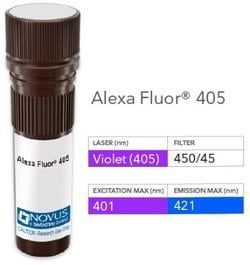
Alexa Fluor 405

Formulation:
50mM Sodium Borate with 0.05% Sodium Azide

Gene Symbols:
PECAM1

Immunogen:
Recombinant full-length human CD31/PECAM-1 protein (Uniprot: P16284)

Quantity:
0.1 mL

Research Discipline:
Angiogenesis, Cancer, Cell Biology, Cellular Markers, Endothelial Cell Markers, Extracellular Matrix, Hematopoietic Stem Cell Markers, Immunology, Mesenchymal Stem Cell Markers, Myeloid Cell Markers, Myeloid derived Suppressor Cell, Signal Transduction, Stem Cell Markers

Test Specificity:
CD31 (PECAM-1) is a transmembrane glycoprotein member of the immunoglobulin supergene family of adhesion molecules. CD31 is expressed by stem cells of the hematopoietic system and is primarily used to identify and concentrate these cells for experimental studies as well as for bone marrow transplantation. Anti-CD31 has shown to be highly specific and sensitive for vascular endothelial cells. Staining of nonvascular tumors (excluding hematopoietic neoplasms) is rare. CD31 monoclonal antibody reacts with normal, benign, and malignant endothelial cells which make up blood vessel lining. The level of CD31 expression can help to determine the degree of tumor angiogenesis, and a high level of CD31 expression may imply a rapidly growing tumor and potentially a predictor of tumor recurrence.

Content And Storage:
Store at 4°C in the dark.

Applications:
Western Blot, Flow Cytometry, Immunohistochemistry, Immunohistochemistry (Paraffin), Immunofluorescence

Clone:
C31.3 + C31.7 + C31.10

Dilution:
Western Blot, Flow Cytometry, Immunohistochemistry, Immunohistochemistry-Paraffin, Immunofluorescence

Gene Alias:
adhesion molecule, CD31, CD31 antigen, CD31/EndoCAM, EndoCAM, FLJ34100, FLJ58394, GPIIA′, PECA1, PECAM-1, PECAM-1, CD31/EndoCAM, platelet endothelial cell adhesion molecule, platelet endothelial cell adhesion molecule-1, platelet/endothelial cell adhesion molecule

Host Species:
Mouse

Purification Method:
Protein A or G purified

Regulatory Status:
RUO

Primary or Secondary:
Primary

Target Species:
Human, Rat, Cynomolgus Monkey, Rabbit

Isotype:
IgG1 κ

Img

Novus Biologicals

NB007354

--


Antigen:
CD31/PECAM-1

Classification:
Monoclonal

Conjugate:
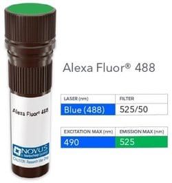
Alexa Fluor 488

Formulation:
50mM Sodium Borate with 0.05% Sodium Azide

Gene Symbols:
PECAM1

Immunogen:
Recombinant full-length human CD31/PECAM-1 protein (Uniprot: P16284)

Quantity:
0.1 mL

Research Discipline:
Angiogenesis, Cancer, Cell Biology, Cellular Markers, Endothelial Cell Markers, Extracellular Matrix, Hematopoietic Stem Cell Markers, Immunology, Mesenchymal Stem Cell Markers, Myeloid Cell Markers, Myeloid derived Suppressor Cell, Signal Transduction, Stem Cell Markers

Test Specificity:
CD31 (PECAM-1) is a transmembrane glycoprotein member of the immunoglobulin supergene family of adhesion molecules. CD31 is expressed by stem cells of the hematopoietic system and is primarily used to identify and concentrate these cells for experimental studies as well as for bone marrow transplantation. Anti-CD31 has shown to be highly specific and sensitive for vascular endothelial cells. Staining of nonvascular tumors (excluding hematopoietic neoplasms) is rare. CD31 monoclonal antibody reacts with normal, benign, and malignant endothelial cells which make up blood vessel lining. The level of CD31 expression can help to determine the degree of tumor angiogenesis, and a high level of CD31 expression may imply a rapidly growing tumor and potentially a predictor of tumor recurrence.

Content And Storage:
Store at 4°C in the dark.

Applications:
Western Blot, Flow Cytometry, Immunohistochemistry, Immunohistochemistry (Paraffin), Immunofluorescence

Clone:
C31.3 + C31.7 + C31.10

Dilution:
Western Blot, Flow Cytometry, Immunohistochemistry, Immunohistochemistry-Paraffin, Immunofluorescence

Gene Alias:
adhesion molecule, CD31, CD31 antigen, CD31/EndoCAM, EndoCAM, FLJ34100, FLJ58394, GPIIA′, PECA1, PECAM-1, PECAM-1, CD31/EndoCAM, platelet endothelial cell adhesion molecule, platelet endothelial cell adhesion molecule-1, platelet/endothelial cell adhesion molecule

Host Species:
Mouse

Purification Method:
Protein A or G purified

Regulatory Status:
RUO

Primary or Secondary:
Primary

Target Species:
Human, Rat, Cynomolgus Monkey, Rabbit

Isotype:
IgG1 κ