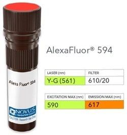
NB007529

CD34 Antibody (HPCA1/763), Alexa Fluor™ 750, Novus Biologicals™

Manufacturer: Novus Biologicals

Select a Size

Pack Size SKU Availability Price
Each of 1 NB007529-Each-of-1 In Stock ₹ 56,298.48

NB007529 - Each of 1

₹ 56,298.48

In Stock

Quantity

1

Base Price: ₹ 56,298.48

GST (18%): ₹ 10,133.726

Total Price: ₹ 66,432.206

Antigen

CD34

Classification

Monoclonal

Conjugate

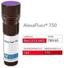
Alexa Fluor 750

Formulation

50mM Sodium Borate with 0.05% Sodium Azide

Gene Symbols

CD34

Immunogen

Recombinant full-length human CD34 protein (Uniprot: P28906)

Quantity

0.1 mL

Research Discipline

Adaptive Immunity, Angiogenesis, B Cell Development and Differentiation Markers, Breast Cancer, Cancer, Cell Biology, Cellular Markers, Endothelial Cell Markers, Hematopoietic Stem Cell Markers, Hypoxia, Immunology, Innate Immunity, Mast Cell Markers, Mesenchymal Stem Cell Markers, Myeloid Cell Markers, Myeloid derived Suppressor Cell, Neuronal Cell Markers, Neuroscience, Stem Cell Markers, Tumor Suppressors

Test Specificity

This monoclonal antibody recognizes a single chain, transmembrane, heavily glycosylated protein of 90-120kDa, which is identified as CD34. Its expression is a hallmark for identifying pluripotent hematopoietic stem or progenitor cells. Its expression is gradually lost as lineage committed progenitors differentiate. CD34 is a marker of choice for staining blasts in acute myeloid leukemia. In addition, CD34 is expressed by soft tissue tumors, such as solitary fibrous tumor and gastrointestinal stromal tumor. Its expression is also found in vascular endothelium. Additionally, it appears that proliferating endothelial cells express this molecule more than the non-proliferating endothelial cells. Anti-CD34 labels 85% of angiosarcoma and Kaposis sarcoma, but with a lower specificity.

Content And Storage

Store at 4°C in the dark.

Applications

Flow Cytometry, Immunohistochemistry, Immunohistochemistry (Paraffin), Immunofluorescence

Clone

HPCA1/763

Dilution

Flow Cytometry, Immunohistochemistry, Immunohistochemistry-Paraffin, Immunofluorescence

Gene Alias

CD34 antigenhematopoietic progenitor cell antigen CD34, CD34 molecule

Host Species

Mouse

Purification Method

Protein A or G purified

Regulatory Status

RUO

Primary or Secondary

Primary

Target Species

Human

Isotype

IgG1 λ

Related Products

Img

Novus Biologicals

NB007527

--

Img

Novus Biologicals

NB007511

--

Img

Novus Biologicals

NB007510

--

Img

Novus Biologicals

NB007505

--

Img

Novus Biologicals

NB007506

--

Img

Novus Biologicals

NB007508

--

Img

Novus Biologicals

NB007514

--

Img

Novus Biologicals

NB007523

--

Description

  • CD34 Monoclonal specifically detects CD34 in Human samples
  • It is validated for Immunohistochemistry, Immunohistochemistry-Paraffin, Immunofluorescence.

Compare Similar Items

Show Difference

Img

Novus Biologicals

NB007529

--


Antigen:
CD34

Classification:
Monoclonal

Conjugate:
Alexa Fluor 750

Formulation:
50mM Sodium Borate with 0.05% Sodium Azide

Gene Symbols:
CD34

Immunogen:
Recombinant full-length human CD34 protein (Uniprot: P28906)

Quantity:
0.1 mL

Research Discipline:
Adaptive Immunity, Angiogenesis, B Cell Development and Differentiation Markers, Breast Cancer, Cancer, Cell Biology, Cellular Markers, Endothelial Cell Markers, Hematopoietic Stem Cell Markers, Hypoxia, Immunology, Innate Immunity, Mast Cell Markers, Mesenchymal Stem Cell Markers, Myeloid Cell Markers, Myeloid derived Suppressor Cell, Neuronal Cell Markers, Neuroscience, Stem Cell Markers, Tumor Suppressors

Test Specificity:
This monoclonal antibody recognizes a single chain, transmembrane, heavily glycosylated protein of 90-120kDa, which is identified as CD34. Its expression is a hallmark for identifying pluripotent hematopoietic stem or progenitor cells. Its expression is gradually lost as lineage committed progenitors differentiate. CD34 is a marker of choice for staining blasts in acute myeloid leukemia. In addition, CD34 is expressed by soft tissue tumors, such as solitary fibrous tumor and gastrointestinal stromal tumor. Its expression is also found in vascular endothelium. Additionally, it appears that proliferating endothelial cells express this molecule more than the non-proliferating endothelial cells. Anti-CD34 labels 85% of angiosarcoma and Kaposis sarcoma, but with a lower specificity.

Content And Storage:
Store at 4°C in the dark.

Applications:
Flow Cytometry, Immunohistochemistry, Immunohistochemistry (Paraffin), Immunofluorescence

Clone:
HPCA1/763

Dilution:
Flow Cytometry, Immunohistochemistry, Immunohistochemistry-Paraffin, Immunofluorescence

Gene Alias:
CD34 antigenhematopoietic progenitor cell antigen CD34, CD34 molecule

Host Species:
Mouse

Purification Method:
Protein A or G purified

Regulatory Status:
RUO

Primary or Secondary:
Primary

Target Species:
Human

Isotype:
IgG1 λ

Img

Novus Biologicals

NB007531

--


Antigen:
Tenascin C

Classification:
Monoclonal

Conjugate:
Alexa Fluor 350

Formulation:
50mM Sodium Borate with 0.05% Sodium Azide

Gene Symbols:
TNC

Immunogen:
Human breast carcinoma (Uniprot: P24821)

Quantity:
0.1 mL

Research Discipline:
Breast Cancer, Cancer, Extracellular Matrix, Neuroscience, Prostate Cancer

Test Specificity:
In Western blotting, it reacts with two bands of ∼MW of 210kDa and 300kDa, identified as two isoforms of Tenascin C. Specificity of this monoclonal antibody is validated by sequential immunoprecipitation with a PAb against Tenascin C. Tenascin C is a multifunctional, disulfide-linkedhexameric extracellular matrix glycoprotein expressed in association with mesenchymal epithelial interactions during development and in the neo-vasculature and stroma of undifferentiated tumors. In adults, it is restricted to certain epithelial-stromal interfaces and increases markedly in hyper-proliferative diseases and in stroma of many neoplasms, including gliomas, breast, squamous and lung carcinomas.

Content And Storage:
Store at 4°C in the dark.

Applications:
Flow Cytometry, Immunohistochemistry, Immunohistochemistry (Paraffin), Immunofluorescence

Clone:
T2H5

Dilution:
Flow Cytometry, Immunohistochemistry, Immunohistochemistry-Paraffin, Immunofluorescence

Gene Alias:
150-225, Cytotactin, Glioma-associated-extracellular matrix antigen, GMEM, GP 150-225, Hexabrachion, hexabrachion (tenascin C, cytotactin), hexabrachion (tenascin), HXBcytotactin, JI, MGC167029, Myotendinous antigen, neuronectin, tenascin, tenascin C, Tenascin-C, tenascin-C isoform 14/AD1/16, TN-C, TNGP

Host Species:
Mouse

Purification Method:
Protein A or G purified

Regulatory Status:
RUO

Primary or Secondary:
Primary

Target Species:
Human, Rat (Negative)

Isotype:
IgG1 κ

Img

Novus Biologicals

NB007532

--


Antigen:
Tenascin C

Classification:
Monoclonal

Conjugate:
Alexa Fluor 532

Formulation:
50mM Sodium Borate with 0.05% Sodium Azide

Gene Symbols:
TNC

Immunogen:
Human breast carcinoma (Uniprot: P24821)

Quantity:
0.1 mL

Research Discipline:
Breast Cancer, Cancer, Extracellular Matrix, Neuroscience, Prostate Cancer

Test Specificity:
In Western blotting, it reacts with two bands of ∼MW of 210kDa and 300kDa, identified as two isoforms of Tenascin C. Specificity of this monoclonal antibody is validated by sequential immunoprecipitation with a PAb against Tenascin C. Tenascin C is a multifunctional, disulfide-linkedhexameric extracellular matrix glycoprotein expressed in association with mesenchymal epithelial interactions during development and in the neo-vasculature and stroma of undifferentiated tumors. In adults, it is restricted to certain epithelial-stromal interfaces and increases markedly in hyper-proliferative diseases and in stroma of many neoplasms, including gliomas, breast, squamous and lung carcinomas.

Content And Storage:
Store at 4°C in the dark.

Applications:
Flow Cytometry, Immunohistochemistry, Immunohistochemistry (Paraffin), Immunofluorescence

Clone:
T2H5

Dilution:
Flow Cytometry, Immunohistochemistry, Immunohistochemistry-Paraffin, Immunofluorescence

Gene Alias:
150-225, Cytotactin, Glioma-associated-extracellular matrix antigen, GMEM, GP 150-225, Hexabrachion, hexabrachion (tenascin C, cytotactin), hexabrachion (tenascin), HXBcytotactin, JI, MGC167029, Myotendinous antigen, neuronectin, tenascin, tenascin C, Tenascin-C, tenascin-C isoform 14/AD1/16, TN-C, TNGP

Host Species:
Mouse

Purification Method:
Protein A or G purified

Regulatory Status:
RUO

Primary or Secondary:
Primary

Target Species:
Human, Rat (Negative)

Isotype:
IgG1 κ

Img

Novus Biologicals

NB007534

--


Antigen:
Tenascin C

Classification:
Monoclonal

Conjugate:
Alexa Fluor 750

Formulation:
50mM Sodium Borate with 0.05% Sodium Azide

Gene Symbols:
TNC

Immunogen:
Human breast carcinoma (Uniprot: P24821)

Quantity:
0.1 mL

Research Discipline:
Breast Cancer, Cancer, Extracellular Matrix, Neuroscience, Prostate Cancer

Test Specificity:
In Western blotting, it reacts with two bands of ∼MW of 210kDa and 300kDa, identified as two isoforms of Tenascin C. Specificity of this monoclonal antibody is validated by sequential immunoprecipitation with a PAb against Tenascin C. Tenascin C is a multifunctional, disulfide-linkedhexameric extracellular matrix glycoprotein expressed in association with mesenchymal epithelial interactions during development and in the neo-vasculature and stroma of undifferentiated tumors. In adults, it is restricted to certain epithelial-stromal interfaces and increases markedly in hyper-proliferative diseases and in stroma of many neoplasms, including gliomas, breast, squamous and lung carcinomas.

Content And Storage:
Store at 4°C in the dark.

Applications:
Flow Cytometry, Immunohistochemistry, Immunohistochemistry (Paraffin), Immunofluorescence

Clone:
T2H5

Dilution:
Flow Cytometry, Immunohistochemistry, Immunohistochemistry-Paraffin, Immunofluorescence

Gene Alias:
150-225, Cytotactin, Glioma-associated-extracellular matrix antigen, GMEM, GP 150-225, Hexabrachion, hexabrachion (tenascin C, cytotactin), hexabrachion (tenascin), HXBcytotactin, JI, MGC167029, Myotendinous antigen, neuronectin, tenascin, tenascin C, Tenascin-C, tenascin-C isoform 14/AD1/16, TN-C, TNGP

Host Species:
Mouse

Purification Method:
Protein A or G purified

Regulatory Status:
RUO

Primary or Secondary:
Primary

Target Species:
Human, Rat (Negative)

Isotype:
IgG1 κ