NB007654

ETS1 associated protein II Antibody (TDP2/1258), PE, Novus Biologicals™

Manufacturer: Novus Biologicals

Select a Size

Pack Size SKU Availability Price
Each of 1 NB007654-Each-of-1 In Stock ₹ 55,271.76

NB007654 - Each of 1

₹ 55,271.76

In Stock

Quantity

1

Base Price: ₹ 55,271.76

GST (18%): ₹ 9,948.917

Total Price: ₹ 65,220.677

Antigen

ETS1 associated protein II

Classification

Monoclonal

Conjugate

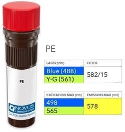
PE

Formulation

PBS with 0.05% Sodium Azide

Gene Symbols

TDP2

Immunogen

Recombinant full-length human ETS1 associated protein II protein (Uniprot: O95551)

Quantity

0.1 mL

Research Discipline

Signal Transduction, Transcription Factors and Regulators

Test Specificity

This MAb recognizes a protein of 41kDa, which is identified as TDP2. It is a member of a superfamily of divalent cation-dependent phosphodiesterases. The encoded protein associates with CD40, tumor necrosis factor (TNF) receptor-75 and TNF receptor associated factors (TRAFs), and inhibits nuclear factor-kappa-B activation. This protein has sequence and structural similarities with APE1 endonuclease, which is involved in both DNA repair and the activation of transcription factors. DNA repair enzyme that can remove a variety of covalent adducts from DNA through hydrolysis of a 5'-phosphodiester bond, giving rise to DNA with a free 5' phosphate. Catalyzes the hydrolysis of dead-end complexes between DNA and the topoisomerase 2 (TOP2) active site tyrosine residue. Hydrolyzes 5'-phosphoglycolates on protruding 5' ends on DNA double-strand breaks (DSBs) due to DNA damage by radiation and free radicals. The 5'-tyrosyl DNA phosphodiesterase activity can enable the repair of TOP2-induced DSBs without the need for nuclease activity, creating a 'clean' DSB with 5'-phosphate termini that are ready for ligation. Has also 3'-tyrosyl DNA phosphodiesterase activity, but less efficiently and much slower than TDP1. May also act as a negative regulator of ETS1 and may inhibit nuclear factor-kappa-B activation.

Content And Storage

Store at 4°C in the dark.

Applications

Flow Cytometry

Clone

TDP2/1258

Dilution

Flow Cytometry

Gene Alias

AD022, dJ30M3.3, EAP2, EAPII, EC 3.1.4.-, ETS1-associated protein 2, ETS1-associated protein II, MGC111021,5'-Tyr-DNA phosphodiesterase, MGC9099,5'-tyrosyl-DNA phosphodiesterase, TRAF and TNF receptor associated protein, TTRAPTRAF and TNF receptor-associated protein, Tyr-DNA phosphodiesterase 2, tyrosyl-DNA phosphodiesterase 2

Host Species

Mouse

Purification Method

Protein A or G purified

Regulatory Status

RUO

Primary or Secondary

Primary

Target Species

Human

Isotype

IgG2b κ

Related Products

Img

Novus Biologicals

NB007653

--

Img

Novus Biologicals

NB007642

--

Img

Novus Biologicals

NB007645

--

Img

Novus Biologicals

NB007634

--

Img

Novus Biologicals

NB007635

--

Img

Novus Biologicals

NB007638

--

Img

Novus Biologicals

NB007639

--

Img

Novus Biologicals

NB007643

--

Description

  • ETS1 associated protein II Monoclonal specifically detects ETS1 associated protein II in Human samples
  • It is validated for Flow Cytometry.

Compare Similar Items

Show Difference

Img

Novus Biologicals

NB007654

--


Antigen:
ETS1 associated protein II

Classification:
Monoclonal

Conjugate:
PE

Formulation:
PBS with 0.05% Sodium Azide

Gene Symbols:
TDP2

Immunogen:
Recombinant full-length human ETS1 associated protein II protein (Uniprot: O95551)

Quantity:
0.1 mL

Research Discipline:
Signal Transduction, Transcription Factors and Regulators

Test Specificity:
This MAb recognizes a protein of 41kDa, which is identified as TDP2. It is a member of a superfamily of divalent cation-dependent phosphodiesterases. The encoded protein associates with CD40, tumor necrosis factor (TNF) receptor-75 and TNF receptor associated factors (TRAFs), and inhibits nuclear factor-kappa-B activation. This protein has sequence and structural similarities with APE1 endonuclease, which is involved in both DNA repair and the activation of transcription factors. DNA repair enzyme that can remove a variety of covalent adducts from DNA through hydrolysis of a 5'-phosphodiester bond, giving rise to DNA with a free 5' phosphate. Catalyzes the hydrolysis of dead-end complexes between DNA and the topoisomerase 2 (TOP2) active site tyrosine residue. Hydrolyzes 5'-phosphoglycolates on protruding 5' ends on DNA double-strand breaks (DSBs) due to DNA damage by radiation and free radicals. The 5'-tyrosyl DNA phosphodiesterase activity can enable the repair of TOP2-induced DSBs without the need for nuclease activity, creating a 'clean' DSB with 5'-phosphate termini that are ready for ligation. Has also 3'-tyrosyl DNA phosphodiesterase activity, but less efficiently and much slower than TDP1. May also act as a negative regulator of ETS1 and may inhibit nuclear factor-kappa-B activation.

Content And Storage:
Store at 4°C in the dark.

Applications:
Flow Cytometry

Clone:
TDP2/1258

Dilution:
Flow Cytometry

Gene Alias:
AD022, dJ30M3.3, EAP2, EAPII, EC 3.1.4.-, ETS1-associated protein 2, ETS1-associated protein II, MGC111021,5'-Tyr-DNA phosphodiesterase, MGC9099,5'-tyrosyl-DNA phosphodiesterase, TRAF and TNF receptor associated protein, TTRAPTRAF and TNF receptor-associated protein, Tyr-DNA phosphodiesterase 2, tyrosyl-DNA phosphodiesterase 2

Host Species:
Mouse

Purification Method:
Protein A or G purified

Regulatory Status:
RUO

Primary or Secondary:
Primary

Target Species:
Human

Isotype:
IgG2b κ

Img

Novus Biologicals

NB007657

--


Antigen:
ETS1 associated protein II

Classification:
Monoclonal

Conjugate:
DyLight 405

Formulation:
50mM Sodium Borate with 0.05% Sodium Azide

Gene Symbols:
TDP2

Immunogen:
Recombinant full-length human ETS1 associated protein II protein (Uniprot: O95551)

Quantity:
0.1 mL

Research Discipline:
Signal Transduction, Transcription Factors and Regulators

Test Specificity:
This MAb recognizes a protein of 41kDa, which is identified as TDP2. It is a member of a superfamily of divalent cation-dependent phosphodiesterases. The encoded protein associates with CD40, tumor necrosis factor (TNF) receptor-75 and TNF receptor associated factors (TRAFs), and inhibits nuclear factor-kappa-B activation. This protein has sequence and structural similarities with APE1 endonuclease, which is involved in both DNA repair and the activation of transcription factors. DNA repair enzyme that can remove a variety of covalent adducts from DNA through hydrolysis of a 5'-phosphodiester bond, giving rise to DNA with a free 5' phosphate. Catalyzes the hydrolysis of dead-end complexes between DNA and the topoisomerase 2 (TOP2) active site tyrosine residue. Hydrolyzes 5'-phosphoglycolates on protruding 5' ends on DNA double-strand breaks (DSBs) due to DNA damage by radiation and free radicals. The 5'-tyrosyl DNA phosphodiesterase activity can enable the repair of TOP2-induced DSBs w

Content And Storage:
Store at 4°C in the dark.

Applications:
Flow Cytometry, Immunohistochemistry, Immunohistochemistry (Paraffin), Immunofluorescence

Clone:
TDP2/1258

Dilution:
Flow Cytometry, Immunohistochemistry, Immunohistochemistry-Paraffin, Immunofluorescence

Gene Alias:
AD022, dJ30M3.3, EAP2, EAPII, EC 3.1.4.-, ETS1-associated protein 2, ETS1-associated protein II, MGC111021,5'-Tyr-DNA phosphodiesterase, MGC9099,5'-tyrosyl-DNA phosphodiesterase, TRAF and TNF receptor associated protein, TTRAPTRAF and TNF receptor-associated protein, Tyr-DNA phosphodiesterase 2, tyrosyl-DNA phosphodiesterase 2

Host Species:
Mouse

Purification Method:
Protein A or G purified

Regulatory Status:
RUO

Primary or Secondary:
Primary

Target Species:
Human

Isotype:
IgG2b κ

Img

Novus Biologicals

NB007658

--


Antigen:
Villin 1

Classification:
Monoclonal

Conjugate:
Alexa Fluor 405

Formulation:
50mM Sodium Borate with 0.05% Sodium Azide

Gene Symbols:
VIL1

Immunogen:
Recombinant human Villin 1 fragment (around aa179-311) (exact sequence is proprietary) (Uniprot: P09327)

Quantity:
0.1 mL

Research Discipline:
Cancer, Cell Biology, Cellular Markers

Test Specificity:
Recognizes a protein of 95kDa, which is identified as villin. It is a major constituent in the microvilli, which compose the brush border of epithelial cells forming absorptive surfaces of the intestinal and renal proximal tubular epithelia. Anti-Villin labels the brush border area in the gastrointestinal mucosal epithelium and urogenital tract. Among neoplasms, villin is predominantly expressed in tumors of colorectal origin. Antibody to villin is useful in identifying malignant cells from primary and metastatic colorectal carcinomas. This antibody also labels Merkel cells of the skin.

Content And Storage:
Store at 4°C in the dark.

Applications:
Western Blot, Flow Cytometry, Immunohistochemistry, Immunohistochemistry (Paraffin), Immunofluorescence

Clone:
VIL1/1314

Dilution:
Western Blot, Flow Cytometry, Immunohistochemistry, Immunohistochemistry-Paraffin, Immunofluorescence

Gene Alias:
D2S1471, VIL, villin 1, villin-1

Host Species:
Mouse

Purification Method:
Protein A or G purified

Regulatory Status:
RUO

Primary or Secondary:
Primary

Target Species:
Human

Isotype:
IgG1 κ

Img

Novus Biologicals

NB007659

--


Antigen:
Villin 1

Classification:
Monoclonal

Conjugate:
Alexa Fluor 488

Formulation:
50mM Sodium Borate with 0.05% Sodium Azide

Gene Symbols:
VIL1

Immunogen:
Recombinant human Villin 1 fragment (around aa179-311) (exact sequence is proprietary) (Uniprot: P09327)

Quantity:
0.1 mL

Research Discipline:
Cancer, Cell Biology, Cellular Markers

Test Specificity:
Recognizes a protein of 95kDa, which is identified as villin. It is a major constituent in the microvilli, which compose the brush border of epithelial cells forming absorptive surfaces of the intestinal and renal proximal tubular epithelia. Anti-Villin labels the brush border area in the gastrointestinal mucosal epithelium and urogenital tract. Among neoplasms, villin is predominantly expressed in tumors of colorectal origin. Antibody to villin is useful in identifying malignant cells from primary and metastatic colorectal carcinomas. This antibody also labels Merkel cells of the skin.

Content And Storage:
Store at 4°C in the dark.

Applications:
Western Blot, Flow Cytometry, Immunohistochemistry, Immunohistochemistry (Paraffin), Immunofluorescence

Clone:
VIL1/1314

Dilution:
Western Blot, Flow Cytometry, Immunohistochemistry, Immunohistochemistry-Paraffin, Immunofluorescence

Gene Alias:
D2S1471, VIL, villin 1, villin-1

Host Species:
Mouse

Purification Method:
Protein A or G purified

Regulatory Status:
RUO

Primary or Secondary:
Primary

Target Species:
Human

Isotype:
IgG1 κ