NB008151

CD3 epsilon Antibody (C3e/1308), Alexa Fluor™ 594, Novus Biologicals™

Manufacturer: Novus Biologicals

Select a Size

Pack Size SKU Availability Price
Each of 1 NB008151-Each-of-1 In Stock ₹ 58,608.60

NB008151 - Each of 1

₹ 58,608.60

In Stock

Quantity

1

Base Price: ₹ 58,608.60

GST (18%): ₹ 10,549.548

Total Price: ₹ 69,158.148

Antigen

CD3 epsilon

Classification

Monoclonal

Conjugate

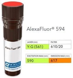
Alexa Fluor 594

Formulation

50mM Sodium Borate with 0.05% Sodium Azide

Gene Symbols

CD3E

Immunogen

Recombinant human CD3 epsilon fragment (aa 23-119) (Uniprot: P07766)

Quantity

0.1 mL

Research Discipline

Adaptive Immunity, Apoptosis, Cytokine Research, Diabetes Research, Immunology, Innate Immunity, Signal Transduction, Stem Cell Lines, Stem Cell Markers

Test Specificity

Recognizes the epsilon-chain of CD3, which consists of five different polypeptide chains (designated as gamma, delta, epsilon, zeta, and eta) with MW ranging from 16-28kDa. The CD3 complex is closely associated at the lymphocyte cell surface with the T cell antigen receptor (TCR). Reportedly, CD3 complex is involved in signal transduction to the T cell interior following antigen recognition. The CD3 antigen is first detectable in early thymocytes and probably represents one of the earliest signs of commitment to the T cell lineage. In cortical thymocytes, CD3 is predominantly intra-cytoplasmic. However, in medullary thymocytes, it appears on the T cell surface. CD3 antigen is a highly specific marker for T cells, and is present in majority of T cell neoplasms.

Content And Storage

Store at 4°C in the dark.

Applications

Western Blot, Flow Cytometry, Immunohistochemistry, Immunocytochemistry, Immunofluorescence, Immunohistochemistry (Paraffin)

Clone

C3e/1308

Dilution

Western Blot, Flow Cytometry, Immunohistochemistry, Immunocytochemistry/Immunofluorescence, Immunohistochemistry-Paraffin, Flow (Intracellular)

Gene Alias

CD3e antigen, CD3e antigen, epsilon polypeptide (TiT3 complex), CD3e molecule, epsilon (CD3-TCR complex), CD3-epsilon, FLJ18683, T3E, T-cell antigen receptor complex, epsilon subunit of T3, T-cell surface antigen T3/Leu-4 epsilon chain, T-cell surface glycoprotein CD3 epsilon chain, TCRE

Host Species

Mouse

Purification Method

Protein A or G purified

Regulatory Status

RUO

Primary or Secondary

Primary

Target Species

Human, Mouse

Isotype

IgG2b κ

Related Products

Img

Novus Biologicals

NB008150

--

Img

Novus Biologicals

NB008149

--

Img

Novus Biologicals

NB008152

--

Img

Novus Biologicals

NB008153

--

Img

Novus Biologicals

NB008173

--

Img

Novus Biologicals

NB008172

--

Img

Novus Biologicals

NB008176

--

Img

Novus Biologicals

NB008018

--

Description

  • CD3 epsilon Monoclonal specifically detects CD3 epsilon in Human, Mouse samples
  • It is validated for Western Blot, Flow Cytometry, Immunohistochemistry, Immunohistochemistry-Paraffin, Flow (Intracellular), Multiplex Immunoassay.

Compare Similar Items

Show Difference

Img

Novus Biologicals

NB008151

--


Antigen:
CD3 epsilon

Classification:
Monoclonal

Conjugate:
Alexa Fluor 594

Formulation:
50mM Sodium Borate with 0.05% Sodium Azide

Gene Symbols:
CD3E

Immunogen:
Recombinant human CD3 epsilon fragment (aa 23-119) (Uniprot: P07766)

Quantity:
0.1 mL

Research Discipline:
Adaptive Immunity, Apoptosis, Cytokine Research, Diabetes Research, Immunology, Innate Immunity, Signal Transduction, Stem Cell Lines, Stem Cell Markers

Test Specificity:
Recognizes the epsilon-chain of CD3, which consists of five different polypeptide chains (designated as gamma, delta, epsilon, zeta, and eta) with MW ranging from 16-28kDa. The CD3 complex is closely associated at the lymphocyte cell surface with the T cell antigen receptor (TCR). Reportedly, CD3 complex is involved in signal transduction to the T cell interior following antigen recognition. The CD3 antigen is first detectable in early thymocytes and probably represents one of the earliest signs of commitment to the T cell lineage. In cortical thymocytes, CD3 is predominantly intra-cytoplasmic. However, in medullary thymocytes, it appears on the T cell surface. CD3 antigen is a highly specific marker for T cells, and is present in majority of T cell neoplasms.

Content And Storage:
Store at 4°C in the dark.

Applications:
Western Blot, Flow Cytometry, Immunohistochemistry, Immunocytochemistry, Immunofluorescence, Immunohistochemistry (Paraffin)

Clone:
C3e/1308

Dilution:
Western Blot, Flow Cytometry, Immunohistochemistry, Immunocytochemistry/Immunofluorescence, Immunohistochemistry-Paraffin, Flow (Intracellular)

Gene Alias:
CD3e antigen, CD3e antigen, epsilon polypeptide (TiT3 complex), CD3e molecule, epsilon (CD3-TCR complex), CD3-epsilon, FLJ18683, T3E, T-cell antigen receptor complex, epsilon subunit of T3, T-cell surface antigen T3/Leu-4 epsilon chain, T-cell surface glycoprotein CD3 epsilon chain, TCRE

Host Species:
Mouse

Purification Method:
Protein A or G purified

Regulatory Status:
RUO

Primary or Secondary:
Primary

Target Species:
Human, Mouse

Isotype:
IgG2b κ

Img

Novus Biologicals

NB008152

--


Antigen:
CD3 epsilon

Classification:
Monoclonal

Conjugate:
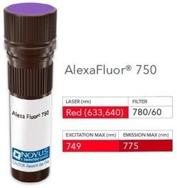
Alexa Fluor 750

Formulation:
50mM Sodium Borate with 0.05% Sodium Azide

Gene Symbols:
CD3E

Immunogen:
Recombinant human CD3 epsilon fragment (aa 23-119) (Uniprot: P07766)

Quantity:
0.1 mL

Research Discipline:
Adaptive Immunity, Apoptosis, Cytokine Research, Diabetes Research, Immunology, Innate Immunity, Signal Transduction, Stem Cell Lines, Stem Cell Markers

Test Specificity:
Recognizes the epsilon-chain of CD3, which consists of five different polypeptide chains (designated as gamma, delta, epsilon, zeta, and eta) with MW ranging from 16-28kDa. The CD3 complex is closely associated at the lymphocyte cell surface with the T cell antigen receptor (TCR). Reportedly, CD3 complex is involved in signal transduction to the T cell interior following antigen recognition. The CD3 antigen is first detectable in early thymocytes and probably represents one of the earliest signs of commitment to the T cell lineage. In cortical thymocytes, CD3 is predominantly intra-cytoplasmic. However, in medullary thymocytes, it appears on the T cell surface. CD3 antigen is a highly specific marker for T cells, and is present in majority of T cell neoplasms.

Content And Storage:
Store at 4°C in the dark.

Applications:
Western Blot, Flow Cytometry, Immunohistochemistry, Immunocytochemistry, Immunofluorescence, Immunohistochemistry (Paraffin)

Clone:
C3e/1308

Dilution:
Western Blot, Flow Cytometry, Immunohistochemistry, Immunocytochemistry/Immunofluorescence, Immunohistochemistry-Paraffin, Flow (Intracellular)

Gene Alias:
CD3e antigen, CD3e antigen, epsilon polypeptide (TiT3 complex), CD3e molecule, epsilon (CD3-TCR complex), CD3-epsilon, FLJ18683, T3E, T-cell antigen receptor complex, epsilon subunit of T3, T-cell surface antigen T3/Leu-4 epsilon chain, T-cell surface glycoprotein CD3 epsilon chain, TCRE

Host Species:
Mouse

Purification Method:
Protein A or G purified

Regulatory Status:
RUO

Primary or Secondary:
Primary

Target Species:
Human, Mouse

Isotype:
IgG2b κ

Img

Novus Biologicals

NB008153

--


Antigen:
CD3 epsilon

Classification:
Monoclonal

Conjugate:
DyLight 488

Formulation:
50mM Sodium Borate with 0.05% Sodium Azide

Gene Symbols:
CD3E

Immunogen:
Recombinant human CD3 epsilon fragment (aa 23-119) (Uniprot: P07766)

Quantity:
0.1 mL

Research Discipline:
Adaptive Immunity, Apoptosis, Cytokine Research, Diabetes Research, Immunology, Innate Immunity, Signal Transduction, Stem Cell Lines, Stem Cell Markers

Test Specificity:
Recognizes the epsilon-chain of CD3, which consists of five different polypeptide chains (designated as gamma, delta, epsilon, zeta, and eta) with MW ranging from 16-28kDa. The CD3 complex is closely associated at the lymphocyte cell surface with the T cell antigen receptor (TCR). Reportedly, CD3 complex is involved in signal transduction to the T cell interior following antigen recognition. The CD3 antigen is first detectable in early thymocytes and probably represents one of the earliest signs of commitment to the T cell lineage. In cortical thymocytes, CD3 is predominantly intra-cytoplasmic. However, in medullary thymocytes, it appears on the T cell surface. CD3 antigen is a highly specific marker for T cells, and is present in majority of T cell neoplasms.

Content And Storage:
Store at 4°C in the dark.

Applications:
Western Blot, Flow Cytometry, Immunohistochemistry, Immunocytochemistry, Immunofluorescence, Immunohistochemistry (Paraffin)

Clone:
C3e/1308

Dilution:
Western Blot, Flow Cytometry, Immunohistochemistry, Immunocytochemistry/Immunofluorescence, Immunohistochemistry-Paraffin, Flow (Intracellular)

Gene Alias:
CD3e antigen, CD3e antigen, epsilon polypeptide (TiT3 complex), CD3e molecule, epsilon (CD3-TCR complex), CD3-epsilon, FLJ18683, T3E, T-cell antigen receptor complex, epsilon subunit of T3, T-cell surface antigen T3/Leu-4 epsilon chain, T-cell surface glycoprotein CD3 epsilon chain, TCRE

Host Species:
Mouse

Purification Method:
Protein A or G purified

Regulatory Status:
RUO

Primary or Secondary:
Primary

Target Species:
Human, Mouse

Isotype:
IgG2b κ

Img

Novus Biologicals

NB008154

--


Antigen:
CD11c

Classification:
Monoclonal

Conjugate:
DyLight 594

Formulation:
50mM Sodium Borate with 0.05% Sodium Azide

Gene Symbols:
ITGAX

Immunogen:
Recombinant human CD11c protein fragment (aa 637-827) (exact sequence is proprietary) (Uniprot: P20702)

Quantity:
0.1 mL

Research Discipline:
Dendritic Cell Markers, Signal Transduction

Test Specificity:
Recognizes a protein of 145kDa, identified as CD11c. CD11c (ITGAX), a member of the leukointegrin family, shares the same beta subunit with other members of the leukocyte adhesion molecule family, which includes CD11a (LFA-1), CD11b (MAC-1) and CD11d (ITGAD), but has a unique alpha chain. CD11c has been shown to play a role in phagocytosis, cell migration, and cytokine production by monocytes/macrophages as well as induction of T-cell proliferation by Langerhans cells. CD11c is expressed prominently on the plasma membranes of monocytes, tissue macrophages, NK cells, and most dendritic cells (DCs). A lower level of expression is also observed on neutrophils as a result of its high level of expression on most DCs. An antibody to CD11c may aid in identification of lesions with histiocytic origin. It may also been used as a marker for hairy cell leukemia in paraffin-embedded tissues.

Content And Storage:
Store at 4°C in the dark.

Applications:
Flow Cytometry, Immunohistochemistry, Immunocytochemistry, Immunofluorescence, Immunohistochemistry (Paraffin)

Clone:
ITGAX/1284

Dilution:
Flow Cytometry, Immunohistochemistry, Immunocytochemistry/Immunofluorescence, Immunohistochemistry-Paraffin

Gene Alias:
95, 95 alpha chain, 95, alpha subunit, CD11 antigen-like family member C, CD11c, CD11c antigen, CD11Cleu M5, alpha subunit, integrin alpha-X, integrin, alpha X (antigen CD11C (p150), alpha polypeptide), integrin, alpha X (complement component 3 receptor 4 subunit), Leu M5, Leukocyte adhesion glycoprotein p150, Leukocyte adhesion receptor p150, leukocyte surface antigen p150, myeloid membrane antigen, alpha subunit, p150 95 integrin alpha chain, SLEB6

Host Species:
Mouse

Purification Method:
Protein A or G purified

Regulatory Status:
RUO

Primary or Secondary:
Primary

Target Species:
Human

Isotype:
IgG2b κ