CD7 Antibody (T3-3A1), PerCP, Novus Biologicals™
Manufacturer: Novus Biologicals
Select a Size
| Pack Size | SKU | Availability | Price |
|---|---|---|---|
| Each of 1 | NB008477-Each-of-1 | In Stock | ₹ 58,562.00 |
NB008477 - Each of 1
In Stock
Quantity
1
Base Price: ₹ 58,562.00
GST (18%): ₹ 10,541.16
Total Price: ₹ 69,103.16
Antigen
CD7
Classification
Monoclonal
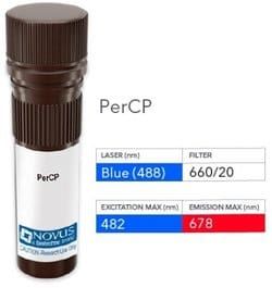
Conjugate
PerCP
Formulation
PBS with 0.05% Sodium Azide
Gene Symbols
CD7
Immunogen
Human T cells
Quantity
0.1 mL
Research Discipline
Cytokine Research, Signal Transduction
Test Specificity
Recognizes a protein of 40kDa, identified as CD7, a member of the immunoglobulin gene superfamily. Its N-terminal amino acids 1-107 are highly homologous to Ig kappa-L chains whereas the carboxyl-terminal region of the extracellular domain is proline-rich and has been postulated to form a stalk from which the Ig domain projects. CD7 is expressed on the majority of immature and mature T-lymphocytes, and T cell leukemia. It is also found on natural killer cells, a small subpopulation of normal B cells and on malignant B cells. Cross-linking surface CD7 positively modulates T cell and NK cell activity as measured by calcium fluxes, expression of adhesion molecules, cytokine secretion and proliferation. CD7 associates directly with phosphoinositol 3'-kinase. CD7 ligation induces production of D-3 phosphoinositides and tyrosine phosphorylation.
Content And Storage
Store at 4°C in the dark.
Applications
Flow Cytometry
Clone
T3-3A1
Dilution
Flow Cytometry
Gene Alias
CD7 antigen, CD7 antigen (p41), CD7 molecule, GP40T-cell surface antigen Leu-9, LEU-9, T-cell antigen CD7, T-cell leukemia antigen, Tp40, TP41p41 protein
Host Species
Mouse
Purification Method
Protein A or G purified
Regulatory Status
RUO
Primary or Secondary
Primary
Target Species
Human
Isotype
IgG1 κ
Related Products


Description
- CD7 Monoclonal specifically detects CD7 in Human samples
- It is validated for Flow Cytometry.
Compare Similar Items
Show Difference
Antigen: CD7
Classification: Monoclonal
Conjugate: PerCP
Formulation: PBS with 0.05% Sodium Azide
Gene Symbols: CD7
Immunogen: Human T cells
Quantity: 0.1 mL
Research Discipline: Cytokine Research, Signal Transduction
Test Specificity: Recognizes a protein of 40kDa, identified as CD7, a member of the immunoglobulin gene superfamily. Its N-terminal amino acids 1-107 are highly homologous to Ig kappa-L chains whereas the carboxyl-terminal region of the extracellular domain is proline-rich and has been postulated to form a stalk from which the Ig domain projects. CD7 is expressed on the majority of immature and mature T-lymphocytes, and T cell leukemia. It is also found on natural killer cells, a small subpopulation of normal B cells and on malignant B cells. Cross-linking surface CD7 positively modulates T cell and NK cell activity as measured by calcium fluxes, expression of adhesion molecules, cytokine secretion and proliferation. CD7 associates directly with phosphoinositol 3'-kinase. CD7 ligation induces production of D-3 phosphoinositides and tyrosine phosphorylation.
Content And Storage: Store at 4°C in the dark.
Applications: Flow Cytometry
Clone: T3-3A1
Dilution: Flow Cytometry
Gene Alias: CD7 antigen, CD7 antigen (p41), CD7 molecule, GP40T-cell surface antigen Leu-9, LEU-9, T-cell antigen CD7, T-cell leukemia antigen, Tp40, TP41p41 protein
Host Species: Mouse
Purification Method: Protein A or G purified
Regulatory Status: RUO
Primary or Secondary: Primary
Target Species: Human
Isotype: IgG1 κ

Antigen: CD7
Classification: Monoclonal
Conjugate: DyLight 350
Formulation: 50mM Sodium Borate with 0.05% Sodium Azide
Gene Symbols: CD7
Immunogen: Human T cells
Quantity: 0.1 mL
Research Discipline: Cytokine Research, Signal Transduction
Test Specificity: Recognizes a protein of 40kDa, identified as CD7, a member of the immunoglobulin gene superfamily. Its N-terminal amino acids 1-107 are highly homologous to Ig kappa-L chains whereas the carboxyl-terminal region of the extracellular domain is proline-rich and has been postulated to form a stalk from which the Ig domain projects. CD7 is expressed on the majority of immature and mature T-lymphocytes, and T cell leukemia. It is also found on natural killer cells, a small subpopulation of normal B cells and on malignant B cells. Cross-linking surface CD7 positively modulates T cell and NK cell activity as measured by calcium fluxes, expression of adhesion molecules, cytokine secretion and proliferation. CD7 associates directly with phosphoinositol 3'-kinase. CD7 ligation induces production of D-3 phosphoinositides and tyrosine phosphorylation.
Content And Storage: Store at 4°C in the dark.
Applications: Flow Cytometry, Immunofluorescence
Clone: T3-3A1
Dilution: Flow Cytometry, Immunofluorescence
Gene Alias: CD7 antigen, CD7 antigen (p41), CD7 molecule, GP40T-cell surface antigen Leu-9, LEU-9, T-cell antigen CD7, T-cell leukemia antigen, Tp40, TP41p41 protein
Host Species: Mouse
Purification Method: Protein A or G purified
Regulatory Status: RUO
Primary or Secondary: Primary
Target Species: Human
Isotype: IgG1 κ
Antigen: CD7
Classification: Monoclonal
Conjugate: DyLight 405
Formulation: 50mM Sodium Borate with 0.05% Sodium Azide
Gene Symbols: CD7
Immunogen: Human T cells
Quantity: 0.1 mL
Research Discipline: Cytokine Research, Signal Transduction
Test Specificity: Recognizes a protein of 40kDa, identified as CD7, a member of the immunoglobulin gene superfamily. Its N-terminal amino acids 1-107 are highly homologous to Ig kappa-L chains whereas the carboxyl-terminal region of the extracellular domain is proline-rich and has been postulated to form a stalk from which the Ig domain projects. CD7 is expressed on the majority of immature and mature T-lymphocytes, and T cell leukemia. It is also found on natural killer cells, a small subpopulation of normal B cells and on malignant B cells. Cross-linking surface CD7 positively modulates T cell and NK cell activity as measured by calcium fluxes, expression of adhesion molecules, cytokine secretion and proliferation. CD7 associates directly with phosphoinositol 3'-kinase. CD7 ligation induces production of D-3 phosphoinositides and tyrosine phosphorylation.
Content And Storage: Store at 4°C in the dark.
Applications: Flow Cytometry, Immunofluorescence
Clone: T3-3A1
Dilution: Flow Cytometry, Immunofluorescence
Gene Alias: CD7 antigen, CD7 antigen (p41), CD7 molecule, GP40T-cell surface antigen Leu-9, LEU-9, T-cell antigen CD7, T-cell leukemia antigen, Tp40, TP41p41 protein
Host Species: Mouse
Purification Method: Protein A or G purified
Regulatory Status: RUO
Primary or Secondary: Primary
Target Species: Human
Isotype: IgG1 κ
